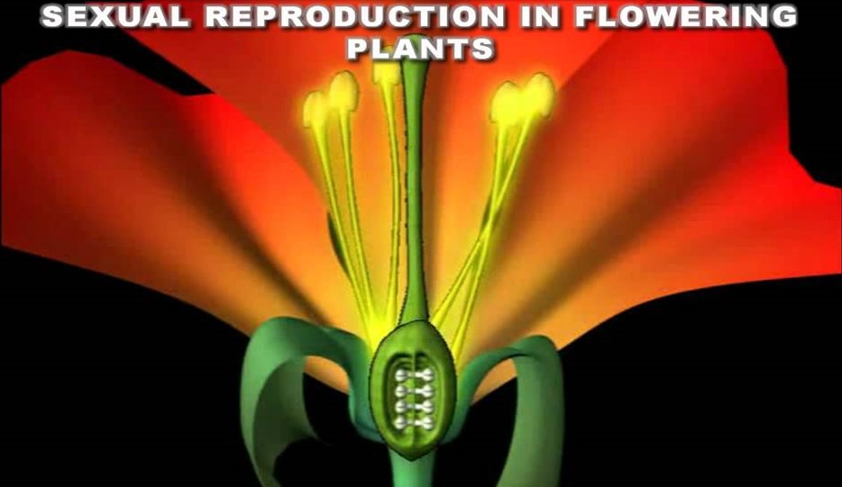
CHAPTER 12 : BIOTECHNOLOGY AND ITS APPLICATIONS

CHAPTER 12

BIOTECHNOLOGY AND ITS APPLICATIONS

  • Biotechnology essentially deals with industrial scale production of biopharmaceuticals and biologicals using genetically modified microbes, fungi, plants and animals.
  • The applications of biotechnology include therapeutics, diagnostics, and genetically
    modified crops for agriculture, processed food, bioremediation, waste treatment, and
    energy production.
  • Three critical research areas of biotechnology are:
    (i) Providing the best catalyst in the form of improved organism usually a microbe or pure enzyme.
    (ii) Creating optimal conditions through engineering for a catalyst to act, and
    (iii) Downstream processing technologies to purify the protein/organic compound.

BIOTECHNOLOGICAL APPLICATIONS IN AGRICULTURE

There are three options that can be thought for increasing food production
(i) agro-chemical based agriculture;
(ii) organic agriculture; and
(iii) Genetically engineered crop-based agriculture.

  •  We have succeeded in tripling the food supply by Green Revolution but yet it was not
    enough to feed the growing human population.
  • Increased yields have partly been due to the use of improved crop varieties, but mainly due to the use of better management practices and use of agrochemicals (fertilisers and pesticides).
  • However, for farmers in the developing world, agrochemicals are often too expensive, and further increases in yield with existing varieties are not possible using conventional breeding.
  • So there is a need to find alternative path that our understanding of genetics can show so that farmers may obtain maximum yield from their fields and to minimise the use of fertilisers and chemicals so that their harmful effects on the environment can be reduced. Use of genetically modified crops is a possible solution.
  • Plants, bacteria, fungi and animals whose genes have been altered by manipulation are called Genetically Modified Organisms (GMO).
  • Genetic modification has:
    (i) Made crops more tolerant to abiotic stresses (cold, drought, salt, heat).
    (ii) Reduced reliance on chemical pesticides (pest-resistant crops).
    (iii) Helped to reduce post-harvest losses.
    (iv) Increased efficiency of mineral usage by plants (this prevents early exhaustion of
    fertility of soil).
    (v) Enhanced nutritional value of food, e.g., Vitamin ‘A’ enriched rice.
    In addition to these uses, GM has been used to create tailor-made plants to supply
    alternative resources to industries, in the form of starches, fuels and pharmaceuticals.
  • By applications of biotechnology in agriculture, pest resistant plants are produced,
    which could decrease the amount of pesticide used.
  • Bt toxin is produced by a bacterium called Bacillus thuringiensis (Bt for short).
  • Bt toxin gene has been cloned from the bacteria and been expressed in plants to
    provide resistance to insects without the need for insecticides; in effect created a
    bio-pesticide. Examples are Bt cotton, Bt corn, rice, tomato, potato and soyabean etc.
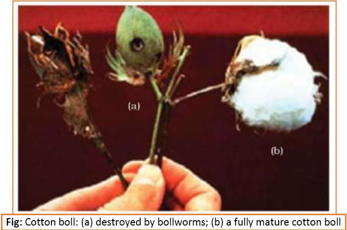
Bt Cotton:

  • Some strains of Bacillus thuringiensis produce proteins that kill certain insects such as lepidopterans (tobacco budworm, armyworm), coleopterans (beetles) and dipterans (flies, mosquitoes).
  • B. thuringiensis forms protein crystals during a particular phase of their growth. These crystals contain a toxic insecticidal protein.
  • This toxin does not kill the Bacillus because this protein exists as inactive protoxins but once an insect ingest the inactive toxin, it is converted into an active form of toxin due to the alkaline pH of the gut which solubilise the crystals. The activated toxin binds to the surface of midgut epithelial cells and create pores that cause cell swelling and lysis and eventually cause death of the insect.
  • Specific Bt toxin genes were isolated from Bacillus thuringiensis and incorporated into the several crop plants such as cotton. The choice of genes depends upon the crop and the targeted pest, as most Bt toxins are insect-group specific.
  • The toxin is coded by a gene named cry. There are a number of them, for example, the proteins encoded by the genes crylAc and cryllAb control the cotton bollworms, that of crylAb controls corn borer.

1

Pest Resistant Plants:

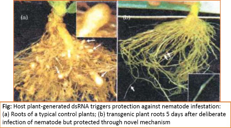
  • Several nematodes parasitise a wide variety of plants and animals including human beings.
  • A nematode Meloidegyne incognitia infects the roots of tobacco plants and causes a
    great reduction in yield.
  • A novel strategy was adopted to prevent this infestation which was based on the
    process of RNA interference (RNAi).
  • RNAi takes place in all eukaryotic organisms as a method of cellular defense.
  • This method involves silencing of a specific mRNA due to a complementary dsRNA
    molecule that binds to and prevents translation of the mRNA (silencing).
  • The source of this complementary RNA could be from an infection by viruses having
    RNA genomes or mobile genetic elements (transposons) that replicate via an RNA
    intermediate.
  • Using Agrobacterium vectors, nematode-specific genes were introduced into the host
    plant.
  • The introduction of DNA was such that it produced both sense and anti-sense RNA in
    the host cells. These two RNA’s being complementary to each other formed a double
    stranded (dsRNA) that initiated RNAi and thus, silenced the specific mRNA of the
    nematode.
  • The consequence was that the parasite could not survive in a transgenic host
    expressing specific interfering RNA. The transgenic plant therefore got itself protected from the parasite.

1

BIOTECHNOLOGICAL APPLICATIONS IN MEDICINE

  • By enabling mass production of safe and more effective therapeutic drugs.
  • Further, the recombinant therapeutics do not induce unwanted immunological
    responses as is common in case of similar products isolated from non-human sources.
  • At present, about 30 recombinant therapeutics have been approved for human-use the world over. In India, 12 of these are presently being marketed.

    Genetically Engineered Insulin

  • Management of adult-onset diabetes is possible by taking insulin at regular time
    intervals.
  • if enough human-insulin was not available, that one would have to isolate and use
    insulin from other animals.
  • Insulin used for diabetes was earlier extracted from pancreas of slaughtered cattle and
    pigs.
  • Insulin from an animal source, though caused some patients to develop allergy or other types of reactions to the foreign protein.
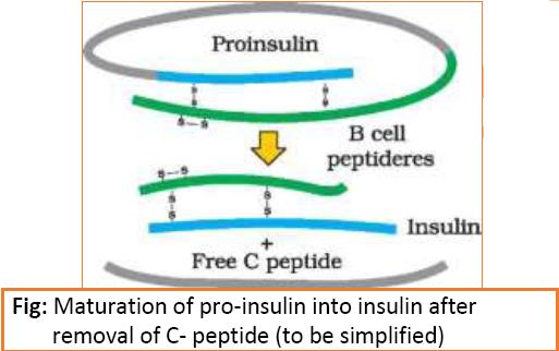
  • Insulin consists of two short polypeptide chains: chain A and chain B, which are linked together by disulphide bridges.
  • In mammals, including humans, insulin is synthesised as a prohormone (like a
    pro-enzyme, the pro-hormone also needs to be processed before it becomes a fully
    mature and functional hormone) which contains an extra stretch called the C peptide.
  • This C peptide is not present in the mature insulin and is removed during maturation
    into insulin.
  • The main challenge for production of insulin using rDNA techniques was getting insulin assembled into a mature form.
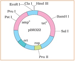
  • In 1983, Eli Lilly an American company prepared two DNA sequences corresponding to A and B, chains of human insulin and introduced them in plasmids of E. coli to produce insulin chains. Chains A and B were produced separately, extracted and combined by creating disulfide bonds to form human insulin.

    1.JPG

Gene Therapy

  • Gene therapy is the corrective therapy for hereditary disease.
    Gene therapy is a collection of methods that allows correction of a gene defect that has
    been diagnosed in a child/embryo. Here genes are inserted into a person’s cells and
    tissues to treat a disease.
  • Correction of a genetic defect involves delivery of a normal gene into the individual or
    embryo to take over the function of and compensate for the non-functional gene.
  • The first clinical gene therapy was given in 1990 to a 4-year old girl with adenosine
    deaminase (ADA) deficiency. This enzyme is crucial for the immune system to function.
  • The disorder is caused due to the deletion of the gene for adenosine deaminase.
  • ADA deficiency can be cured by bone marrow transplantation or by enzyme
    replacement therapy, in which functional ADA is given to the patient by injection.
    But the problem with both of these approaches that they are not completely curative.
  • In gene therapy, lymphocytes from the blood of the patient are grown in a culture
    outside the body. A functional ADA cDNA (using a retroviral vector) is then introduced
    into these lymphocytes, which are subsequently returned to the patient. However, as
    these cells are not immortal, the patient requires periodic infusion of such genetically
    engineered lymphocytes. However, if the gene isolate from marrow cells producing
    ADA is introduced into cells at early embryonic stages, it could be a permanent cure.

Molecular Diagnosis

  • For effective treatment of a disease, early diagnosis and understanding its
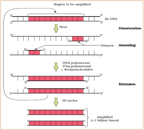
    pathophysiology is very important but using conventional methods of diagnosis (serum and urine analysis, etc.) early detection is not possible.
  • Recombinant DNA technology, Polymerase Chain Reaction (PCR) and Enzyme Linked
    Immuno-sorbent Assay (ELISA) are some of the techniques that serve the purpose of
    early diagnosis.
  • Presence of a pathogen (bacteria, viruses, etc.) is normally suspected only when the
    pathogen has produced a disease symptom. By this time the concentration of pathogen is already very high in the body. However, very low concentration of a bacteria or virus (at a time when the symptoms of the disease are not yet visible) can be detected by amplification of their nucleic acid by PCR.
  • PCR is now routinely used to detect HIV in suspected AIDS patients. It is being used to
    detect mutations in genes in suspected cancer patients too. It is a powerful techqnique
    to identify many other genetic disorders.
  • PCR –
    A single stranded DNA or RNA, tagged with a radioactive molecule (probe) is allowed to hybridise to its complementary DNA in a clone of cells followed by detection using
    autoradiography. The clone having the mutated gene will hence not appear on the
    photographic film, because the probe will not have complimentarity with the mutated
    gene.
  • ELISA is based on the principle of antigen-antibody interaction. Infection by pathogen can be detected by the presence of antigens (proteins, glycoproteins, etc.) or by detecting the antibodies synthesised against the pathogen.

TRANSGENIC ANIMALS

  • Animals that have had their DNA manipulated to possess and express an extra (foreign) gene are known as transgenic animals.
  • Transgenic rats, rabbits, pigs, sheep, cows and fish have been produced, although over 95 per cent of all existing transgenic animals are mice.
  • common reasons to produce transgenic animals:
    (i) Normal physiology and development:
    Transgenic animals can be specifically designed to allow the study of how genes are
    regulated, and how they affect the normal functions of the body and its
    development, e.g., study of complex factors involved in growth such as insulin-like
    growth factor.
    By introducing genes from other species that alter the formation of this factor and
    studying the biological effects that result, information is obtained about the
    biological role of the factor in the body.
    (ii) Study of disease:
    Many transgenic animals are designed to increase our understanding of how genes
    contribute to the development of disease. These are specially made to serve as
    models for human diseases so that Investigation of new treatments for diseases is
    made possible.
    Today transgenic models exist for many human diseases such as cancer, cystic
    fibrosis, rheumatoid arthritis and Alzheimer’s.
    (iii) Biological products:
    Medicines required to treat certain human diseases can contain biological products,
    but such products are often expensive to make.
    Transgenic animals that produce useful biological products can be created by the
    introduction of the portion of DNA (or genes) which codes for a particular product
    such as human protein (α-1-antitrypsin) used to treat emphysema.
    Similar attempts are being made for treatment of phenylketonuria (PKU) and cystic
    fibrosis.
    In 1997, the first transgenic cow, Rosie, produced human protein-enriched milk (2.4
    grams per litre). The milk contained the human alpha-lactalbumin and was
    nutritionally a more balanced product for human babies than natural cow-milk.
    (iv) Vaccine safety:
    Transgenic mice are being developed for use in testing the safety of vaccines before
    they are used on humans.
    Transgenic mice are being used to test the safety of the polio vaccine. If successful
    and found to be reliable, they could replace the use of monkeys to test the safety of
    batches of the vaccine.
    (v) Chemical safety testing:
    This is known as toxicity/safety testing. The procedure is the same as that used for
    testing toxicity of drugs.
    Transgenic animals are made that carry genes which make them more sensitive to
    toxic substances than non-transgenic animals. They are then exposed to the toxic
    substances and the effects studied. Toxicity testing in such animals will allow us to
    obtain results in less time.

    ETHICAL ISSUES

    The manipulation of living organisms by the human race cannot go on any further, without
    regulation. Some ethical standards are required to evaluate the morality of all human
    activities that might help or harm living organisms.
    Going beyond the morality of such issues, the biological significance of such things is also
    important. Genetic modification of organisms can have unpredicatable results when such
    organisms are introduced into the ecosystem.
    Therefore, the Indian Government has set up organisations such as GEAC (Genetic
    Engineering Approval Committee), which will make decisions regarding the validity of GM
    research and the safety of introducing GM-organisms for public services.

Bio-patent:

  • The modification/usage of living organisms for public services (as food and medicine
    sources, for example) has also created problems with patents granted for the same.
  • There is growing public anger that certain companies are being granted patents for
    products and technologies that make use of the genetic materials, plants and other
    biological resources that have long been identified, developed and used by farmers and
    indigenous people of a specific region/country.
  • Rice is an important food grain, the presence of which goes back thousands of years in
    Asia’s agricultural history. There are an estimated 200,000 varieties of rice in India
    alone. The diversity of rice in India is one of the richest in the world.
  • Basmati rice is distinct for its unique aroma and flavour and 27 documented varieties of Basmati are grown in India. There is reference to Basmati in ancient texts, folklore and poetry, as it has been grown for centuries.
  • In 1997, an American company got patent rights on Basmati rice through the US Patent and Trademark Office. This allowed the company to sell a ‘new’ variety of Basmati, in the US and abroad.
  • This ‘new’ variety of Basmati had actually been derived from Indian farmer’s varieties.
    Indian Basmati was crossed with semi-dwarf varieties and claimed as an invention or a novelty.
  • The patent extends to functional equivalents, implying that other people selling
    Basmati rice could be restricted by the patent.
  • Several attempts have also been made to patent uses, products and processes based
    on Indian traditional herbal medicines, e.g., turmeric neem.
  • If we are not vigilant and we do not immediately counter these patent applications,
    other countries/individuals may encash on our rich legacy and we may not be able to
    do anything about it.

Biopiracy

  • It is the term used to refer to the use of bio-resources by multinational companies and
    other organisations without proper authorisation from the countries and people
    concerned without compensatory payment.
  • Most of the industrialised nations are rich financially but poor in biodiversity and
    traditional knowledge. In contrast the developing and the underdeveloped world is rich in biodiversity and traditional knowledge related to bio-resources. Traditional
    knowledge related to bio-resources can be exploited to develop modern applications
    and can also be used to save time, effort and expenditure during their
    commercialisation.
  • There has been growing realisation of the injustice, inadequate compensation and
    benefit sharing between developed and developing countries. Therefore, some nations
    are developing laws to prevent such unauthorised exploitation of their bio-resources
    and traditional knowledge.
  • The Indian Parliament has recently cleared the second amendment of the Indian
    Patents Bill, that takes such issues into consideration, including patent terms
    emergency provisions and research and development initiative.


To download notes in pdf format please click on the following link.

CHAPTER 12 : BIOTECHNOLOGY AND ITS APPLICATIONS

CHAPTER 11 : BIOTECHNOLOGY: PRINCIPLES AND PROCESSES

Chapter 11

Biotechnology : Principles and Processes

[you can download the notes from the link given at the end of theory]

Biotechnology deals with techniques of using live organisms or enzymes from organisms to produce products and processes useful to humans.

  • Traditional form – based on natural capabilities of microorganisms. making curd, bread or wine, which are all microbe-mediated processes, could also be thought as a form of biotechnology. However, it is used in a restricted sense today,
  • Modern form – it uses genetically modified organisms to achieve the same on a larger scale. Further, many other processes/techniques are also included under biotechnology. For example, in vitro fertilisation leading to a ‘test-tube’ baby, synthesising a gene and using it, developing a DNA vaccine or correcting a defective gene, are all part of biotechnology.
  • The European Federation of Biotechnology (EFB) has given a definition of biotechnology that encompasses both traditional view and modern molecular biotechnology. The definition given by EFB is as follows:

‘The integration of natural science and organisms, cells, parts thereof, and molecular analogues for products and services’.

PRINCIPLES OF BIOTECHNOLOGY

  • Among many, the two core techniques that enabled birth of modern biotechnology are :
    • Genetic engineering: Techniques to alter the chemistry of genetic material (DNA and RNA),to introduce these into host organisms and thus change the phenotype of the host organism.
    • Maintenance of sterile (microbial contamination-free) ambience in chemical engineering processes to enable growth of only the desired microbe/eukaryotic cell in large quantities for the manufacture of biotechnological products like antibiotics, vaccines, enzymes, etc.
  • Sexual reproduction has many advantages over asexual reproduction. The former provides opportunities for variations and formulation of unique combinations of genetic setup, some of which may be beneficial to the organism as well as the population. Asexual reproduction preserves the genetic information, while sexual reproduction permits variation.
  • Traditional hybridisation procedures used in plant and animal breeding, very often lead to inclusion and multiplication of undesirable genes along with the desired genes. The techniques of genetic engineering which include creation of recombinant DNA, use of gene cloning and gene transfer, overcome this limitation and allow us to isolate and introduce only one or a set of desirable genes without introducing undesirable genes into the target organism.
  • A piece of DNA, which is somehow transferred into an alien organism, most likely would not be able to multiply itself in the progeny cells of the organism. But, when it gets integrated into the genome of the recipient, it may multiply and be inherited along with the host DNA. This is because the alien piece of DNA has become part of a chromosome, which has the ability to replicate.
  • In a chromosome there is a specific DNA sequence called the origin of replication, which is responsible for initiating replication. Therefore, for the multiplication of any alien piece of DNA in an organism it needs to be a part of a chromosome(s) which has a specific sequence known as ‘origin of replication’. Thus, an alien DNA is linked with the origin of replication, so that, this alien piece of DNA can replicate and multiply itself in the host organism. This can also be called as cloning or making multiple identical copies of any template DNA.
  • The construction of the first recombinant DNA emerged from the possibility of linking a gene encoding antibiotic resistance with a native plasmid (autonomously replicating circular extra-chromosomal DNA) of  Salmonella typhimurium.
  • Stanley Cohen and Herbert Boyer accomplished this in 1972 by isolating the antibiotic resistance gene by cutting out a piece of DNA from a plasmid which was responsible for conferring antibiotic resistance.
  • The cutting of DNA at specific locations became possible with the discovery of the so-called ‘molecular scissors’- restriction enzymes.
  • The cut piece of DNA was then linked with the plasmid DNA. These plasmid DNA act as vectors to transfer the piece of DNA attached to it. A plasmid can be used as vector to deliver an alien piece of DNA into the host organism.
  • The linking of antibiotic resistance gene with the plasmid vector became possible with the enzyme DNA ligase, which acts on cut DNA molecules and joins their ends. This makes a new combination of circular autonomously replicating DNA created in vitro and is known as recombinant DNA.
  • When this DNA is transferred into Escherichia coli, a bacterium closely related to Salmonella, it could replicate using the new host’s DNA polymerase enzyme and make multiple copies. The ability to multiply copies of antibiotic resistance gene in coli was called cloning of antibiotic resistance gene in E. coli.
  • there are three basic steps in genetically modifying an organism
    • identification of DNA with desirable genes;
    • introduction of the identified DNA into the host;
    • maintenance of introduced DNA in the host and transfer of the DNA to its progeny.

TOOLS OF RECOMBINANT DNA TECHNOLOGY

Key tools of Recombinant DNA technology are – restriction enzymes, polymerase enzymes, ligases, vectors and the host organism.

  1. Restriction Enzymes

  • In 1963, the two enzymes responsible for restricting the growth of bacteriophage in Escherichia coli were isolated. One of these added methyl groups to DNA, while the other cut DNA. The later was called restriction endonuclease.
  • The first restriction endonuclease isolated – Hind II.
  • Restriction endonuclease cut DNA molecules at a particular point by recognising a specific sequence of base pairs. This specific base sequence is known as the recognition sequence.(For Hind II – sequence of 6 base pairs).
  • Today we know more than 900 restriction enzymes that have been isolated from over 230 strains of bacteria each of which recognise different recognition sequences.

Naming of enzymes –

  • First letter of the name comes from the genes
  • The second two letters come from the species of the prokaryotic cell from which they were isolated, e.g., EcoRI comes from Escherichia coli RY 13.
  • Next letter derived from the name of strain.
  • Roman numbers following the names indicate the order in which the enzymes were isolated from that strain of bacteria.

Action of enzyme –

  • Restriction enzymes belong to a larger class of enzymes called nucleases. These are of two kinds; exonucleasesand endonucleases.
  • Exonucleases remove nucleotides from the ends of the DNA whereas, endonucleases make cuts at specific positions within the DNA.
  • Each restriction endonuclease functions by ‘inspecting’ the length of a DNA sequence. Once it finds its specific recognition sequence, it will bind to the DNA and cut each of the two strands of the double helix at specific points in their sugar -phosphate backbones.
  • Each restriction endonuclease recognises a specific palindromic nucleotide sequences in the DNA.
  • The palindrome in DNA is a sequence of base pairs that reads same on the two strands when orientation of reading is kept the same. For example, the following sequences reads the same on the two strands in 5→3 This is also true if read in the 3→5direction.

5—— GAATTC —— 3

3—— CTTAAG —— 5

  • Restriction enzymes cut the strand of DNA a little away from the centre of the palindrome sites, but between the same two bases on the opposite strands. This leaves single stranded portions at the ends. There are overhanging stretches called sticky ends on each strand.
  • These are named so because they form hydrogen bonds with their complementary cut counterparts. This stickiness of the ends facilitates the action of the enzyme DNA ligase.
  • Restriction endonucleases are used in genetic engineering to form ‘recombinant’ molecules of DNA, which are composed of DNA from different sources/genomes.
  • When cut by the same restriction enzyme, the resultant DNA fragments have the same kind of ‘sticky-ends’ and, these can be joined together (end-to-end) using DNA ligases .
  • Normally, unless one cuts the vector and the source DNA with the same restriction enzyme, the recombinant vector molecule cannot be created.

1.jpg

Fig: Steps in formation of recombinant DNA by action of restriction endonuclease enzyme – EcoRI

1.jpg

Fig: Diagrammatic representation of recombinant DNA technology


Separation and isolation of DNA fragments :

  • The cutting of DNA by restriction endonucleases results in the fragmentes of DNA. These fragments can be separated by a technique known as gel electrophoresis.
  • Since DNA fragments are negatively charged molecules they can be separated by forcing them to move towards the anode under an electric field through a medium/matrix. Nowadays the most commonly used matrix is agarose which is a natural polymer extracted from sea weeds.
  • The DNA fragments separate (resolve) according to their size through sieving effect provided by the agarose gel. Hence, the smaller the fragment size, the farther it moves.
  • The separated DNA fragments can be visualised only after staining the DNA with a compound known as ethidium bromide followed by exposure to UV radiation.
  • We can see bright orange coloured bands of DNA in aethidium bromide stained gel exposed to UV light.
  • The separated bands of DNA are cut out from the agarose gel and extracted from the gel piece. This step is known as elution. The DNA fragments purified in this way are used in constructing recombinant DNA by joining them with cloning vectors.

1.jpg

Fig: A typical agarose gel electrophoresis showing migration of undigested (lane 1) and digested set of DNA fragments (lane 2 to 4)
  1. Cloning Vectors

  • Plasmids and bacteriophages have the ability to replicate within bacterial cells independent of the control of chromosomal DNA.
  • Bacteriophages because of their high number per cell, have very high copy numbers of their genome within the bacterial cells.
  • If we are able to link an alien piece of DNA with bacteriophage or plasmid DNA, we can multiply its numbers equal to the copy number of the plasmid or bacteriophage.
  • Vectors used at present, are engineered in such way that they help easy linking of foreign DNA and selection of recombinants from non-recombinants.

Features required to facilitate cloning into a vector.

Origin of replication (ori):

  • This is a sequence from where replication starts and any piece of DNA when linked to this sequence can be made to replicate within the host cells.
  • This sequence is also responsible for controlling the copy number of the linked DNA.
  • So, if one wants to recover many copies of the target DNA it should be cloned in a vector whose origin support high copy number.

    Selectable marker :

  • In addition to ‘ori’, the vector requires a selectable marker, which helps in identifying and eliminating nontransformants and selectively permitting the growth of the transformants.
  • Transformation is a procedure through which a piece of DNA is introduced in a host bacterium.
  • Normally, the genes encoding resistance to antibiotics such as ampicillin, chloramphenicol, tetracycline or kanamycin, etc., are considered useful selectable markers for coli. The normal E. coli cells do not carry resistance against any of these antibiotics.

    Cloning sites:

  • In order to link the alien DNA, the vector needs to have very few, preferably single, recognition sites for the commonly used restriction enzymes.
  • Presence of more than one recognition sites within the vector will generate several fragments, which will complicate the gene cloning.
  • The ligation of alien DNA is carried out at a restriction site present in one of the two antibiotic resistance
  • For example, you can ligate a foreign DNA at the Bam H I site of tetracycline resistance gene in the vector pBR322. The recombinant plasmids will lose tetracycline resistance due to insertion of foreign DNA but can still be selected out from non-recombinant ones by plating the transformants on ampicillin containing medium. The transformants growing on ampicillin containing medium are then transferred on a medium containing tetracycline. The recombinants will grow in ampicillin containing medium but not on that containing tetracycline. But, nonrecombinants will grow on the medium containing both the antibiotics. In this case, one antibiotic resistance gene helps in selecting the transformants, whereas the other antibiotic resistance gene gets ‘inactivated due to insertion’ of alien DNA, and helps in selection of recombinants.
  • Selection of recombinants due to inactivation of antibiotics is a cumbersome procedure because it requires simultaneous plating on two plates having different antibiotics. Therefore, alternative selectable markers have been developed which differentiate recombinants from non-recombinants on the basis of their ability to produce colour in the presence of a chromogenic substrate.
  • In this, a recombinant DNA is inserted within the coding sequence of an enzyme, a-galactosidase. This results into inactivation of the enzyme, which is referred to as insertional inactivation. The presence of a chromogenic substrate gives blue coloured colonies if the plasmid in the bacteria does not have an insert. Presence of insert results into insertional inactivation of the a-galactosidase and the colonies do not produce any colour, these are identified as recombinant colonies.

    Vectors for cloning genes in plants and animals :

  • Viruses and bacteria are used to transfer genes into plants and animals which transform eukaryotic cells and force them to do what the bacteria or viruses want.
  • For example, Agrobacterioumtumifaciens, a pathogen of several dicot plants is able to deliver a piece of DNA known as ‘T-DNA’ to transform normal plant cells into a tumor and direct these tumor cells to produce the chemicals required by the pathogen.
  • Similarly, retroviruses in animals have the ability to transform normal cells into cancerous
  • A better understanding of the art of delivering genes by pathogens in their eukaryotic hosts has generated knowledge to transform these tools of pathogens into useful vectors for delivering genes of interest to humans.
  • The tumor inducing (Ti) plasmid of Agrobacterium tumifacienshas now been modified into a cloning vector which is no more pathogenic to the plants but is still able to use the mechanisms to deliver genes of our interest into a variety of plants. Similarly, retroviruses have also been disarmed and are now used to deliver desirable genes into animal cells. So, once a gene or a DNA fragment has been ligated into a suitable vector it is transferred into a bacterial, plant or animal host (where it multiplies).
    1.jpg
Fig: E. coli cloning vector pBR322 showing restriction sites (Hind III, EcoR I, BamH I, Sal I, PvuII, PstI, ClaI), ori and antibiotic resistance genes (ampR and tetR). Rop codes for the proteins involved in the replication of the plasmid.
  1. Competent Host (For Transformation with Recombinant DNA)

  • Since DNA is a hydrophilic molecule, it cannot pass through cell membranes. In order to force bacteria to take up the plasmid, the bacterial cells must first be made ‘competent’ to take up DNA.
  • This is done by treating them with a specific concentration of a divalent cation, such as calcium, which increases the efficiency with which DNA enters the bacterium through pores in its cell wall.
  • Recombinant DNA can then be forced into such cells by incubating the cells with recombinant DNA on ice, followed by placing them briefly at 420oC (heat shock), and then putting them back on ice. This enables the bacteria to take up the recombinant DNA.
  • In micro-injection method, recombinant DNA is directly injected into the nucleus of an animal cell.
  • In another method, suitable for plants, cells are bombarded with high velocity micro-particles of gold or tungsten coated with DNA in a method known as biolisticsor gene gun.
  • And the last method uses ‘disarmed pathogen’ vectors, which when allowed to infect the cell, transfer the recombinant DNA into the host.

PROCESSES OF RECOMBINANT DNA TECHNOLOGY

Recombinant DNA technology involves several steps in specific sequence such as –

  • isolation of DNA,
  • fragmentation of DNA by restriction endonucleases,
  • isolation of a desired DNA fragment,
  • ligation of the DNA fragment into a vector,
  • transferring the recombinant DNA into the host,
  • culturing the host cells in a medium at large scale and
  • extraction of the desired product.
  1. Isolation of the Genetic Material (DNA)

  • Nucleic acid is the genetic material of all organisms without exception. In majority of organisms this is deoxyribonucleic acid or DNA.
  • In order to cut the DNA with restriction enzymes, it needs to be in pure form, free from other macro-molecules. Since the DNA is enclosed within the membranes, we have to break the cell open to release DNA along with other macromolecules such as RNA, proteins, polysaccharides and also lipids. This can be achieved by treating the bacterial cells/plant or animal tissue with enzymes such as lysozyme (bacteria), cellulase(plant cells), chitinase(fungus).
  • genes are located on long molecules of DNA interwined with proteins such as histones.
  • RNA can be removed by treatment with ribonuclease whereas proteins can be removed by treatment with protease. Other molecules can be removed by appropriate treatments and purified DNA ultimately precipitates out after the addition of chilled ethanol. This can be seen as collection of fine threads in the suspension.

1.jpg

Fig: DNA thatseparates out can beremoved by spooling
  1. Cutting of DNA at Specific Locations

  • Restriction enzyme digestions are performed by incubating purified DNA molecules with the restriction enzyme, at the optimal conditions for that specific enzyme.
  • Agarose gel electrophoresis is employed to check the progression of a restriction enzyme digestion. DNA is a negatively charged molecule, hence it moves towards the positive electrode (anode).
  • The process is repeated with the vector DNA also.
  • The joining of DNA involves several processes. After having cut the source DNA as well as the vector DNA with a specific restriction enzyme, the cut out ‘gene of interest’ from the source DNA and the cut vector with space are mixed and ligase is added. This results in the preparation of recombinant DNA.
  1. Amplification of Gene of Interest using PCR (Polymerase Chain Reaction)

  • In this reaction, multiple copies of the gene (or DNA) of interest is synthesisedin vitro using two sets of primers (small chemically synthesised oligonucleotides that are complementary to the regions of DNA) and the enzyme DNA polymerase.
  • The enzyme extends the primers using the nucleotides provided in the reaction and the genomic DNA as template.
  • If the process of replication of DNA is repeated many times, the segment of DNA can be amplified to approximately billion times.
  • Such repeated amplification is achieved by the use of a thermostable DNA polymerase (isolated from a bacterium, Thermusaquaticus), which remain active during the high temperature induced denaturation of double stranded DNA.
  • The amplified fragment if desired can now be used to ligate with a vector for further cloning.

1.jpg

Fig: Polymerase chain reaction (PCR) : Each cycle has three steps: (i) Denaturation;

(ii) Primer annealing; and (iii) Extension of primers

  1. Insertion of Recombinant DNA into the Host Cell/Organism

  • There are several methods of introducing the ligated DNA into recipient cells. Recipient cells after making them ‘competent’ to receive, take up DNA present in its surrounding.
  • So, if a recombinant DNA bearing gene for resistance to an antibiotic (e.g., ampicillin) is transferred into coli cells, the host cells become transformed into ampicillin-resistant cells. If we spread the transformed cells on agar plates containing ampicillin, only transformants will grow, untransformed recipient cells will die. Since, due to ampicillin resistance gene, one is able to select a transformed cell in the presence of ampicillin. The ampicillin resistance gene in this case is called a selectable marker.
  1. Obtaining the Foreign Gene Product

  • When you insert a piece of alien DNA into a cloning vector and transfer it into a bacterial, plant or animal cell, the alien DNA gets multiplied.
  • In almost all recominant technologies, the ultimate aim is to produce a desirable protein. Hence, there is a need for the recombinant DNA to be expressed.
  • The foreign gene gets expressed under appropriate conditions. The expression of foreign genes in host cells involve understanding many technical details.
  • After having cloned the gene of interest and having optimised the conditions to induce the expression of the target protein, one has to consider producing it on a large scale.
  • If any protein encoding gene is expressed in a heterologous host, is called a recombinant protein.
  • The cells harbouring cloned genes of interest may be grown on a small scale in the laboratory. The cultures may be used for extracting the desired protein and then purifying it by using different separation techniques.
  • The cells can also be multiplied in a continuous culture system wherein the used medium is drained out from one side while fresh medium is added from the other to maintain the cells in their physiologically most active log/exponential phase. This type of culturing method produces a larger biomass leading to higher yields of desired protein.
  • Small volume cultures cannot yield appreciable quantities of products. To produce in large quantities, the development of bioreactors, where large volumes (100-1000 litres) of culture can be processed, was required. Thus, bioreactors can be thought of as vessels in which raw materials are biologically converted into specific products, individual enzymes, etc., using microbial plant, animal or human cells. A bioreactor provides the optimal conditions for achieving the desired product by providing optimum growth conditions (temperature, pH, substrate, salts, vitamins, oxygen).
  • A stirred-tank reactor is usually cylindrical or with a curved base to facilitate the mixing of the reactor contents. The stirrer facilitates even mixing and oxygen availability throughout the bioreactor. Alternatively air can be bubbled through the reactor.
  • The bioreactor has an agitator system, an oxygen delivery system and a foam control system, a temperature control system, pH control system and sampling ports so that small volumes of the culture can be withdrawn periodically.

1.jpg

Fig: (a) Simple stirred-tank bioreactor; (b) Sparged stirred-tank bioreactor through whichsterile air bubbles are sparged


  1. Downstream Processing
  • After completion of the biosynthetic stage, the product has to be subjected through a series of processes before it is ready for marketing as a finished The processes include separation and purification, which are collectively referred to as downstream processing.
  • The product has to be formulated with suitable preservatives. Such formulation has to undergo thorough clinical trials as in case of drugs. Strict quality control testing for each product is also required. The downstream processing and quality control testing vary from product to product.

To download notes in pdf format please click on the following link.

CHAPTER 11 : BIOTECHNOLOGY: PRINCIPLES AND PROCESSES

CHAPTER 2 : SEXUAL REPRODUCTION IN FLOWERING PLANTS

CHAPTER 2

SEXUAL REPRODUCTION IN FLOWERING PLANTS

[you can download the notes from the link given at the end of theory]

  • All flowering plants show sexual reproduction.

Flower – A Fascinating Organ of Angiosperms

  • Flowers are objects of aesthetic, ornamental, social, religious and cultural value – they have always been used as symbols for conveying important human feelings such as love, affection, happiness, grief, mourning, etc.

1

  • Pre-fertilisation: Structures and Events

  • Several hormonal and structural changes are initiated which lead to the differentiation and further development of the floral primordium.
  • Inflorescences are formed which bear the floral buds and then the flowers.
  • In the flower the male and female reproductive structures, the androecium and the gynoecium differentiate and develop.
  • The androecium consists of a whorl of stamens representing the male reproductive organ and the gynoecium represents the female reproductive organ.

Stamen, Microsporangium and Pollen Grain

  • A typical stamen has two parts –

the long and slender stalk called the filament,

and the terminal generally bilobed structure called the anther.

  • The proximal end of the filament is attached to the thalamus or the petal of the flower.
  • The number and length of stamens are variable in flowers of different species.
  • A typical angiosperm anther is bilobed with each lobe having two theca, i.e., they are
  • The anther is a four-sided (tetragonal) structure consisting of four microsporangia located at the corners, two in each lobe. tetrasporangiate
  • The microsporangia develop further and become pollen sacs. They extend longitudinally all through the length of an anther and are packed with pollen grains.

1

1.jpg

Structure of microsporangium:

  • In a transverse of an anther section, a typical microsporangium appears nearcircular in outline.
  • It is generally surrounded by four wall layers :- the epidermis, endothecium, middle layers and the tapetum.
  • The outer three wall layers perform the function of protection and help in dehiscence of anther to release the pollen.
  • The innermost wall layer is the It nourishes the developing pollen grains. Cells of the tapetum possess dense cytoplasm and generally have more than one nucleus.
  • When the anther is young, a group of compactly arranged homogenous cells called the sporogenous tissue occupies the centre of each microsporangium.

Microsporogenesis :

  • As the anther develops, the cells of the sporogenous tissue undergo meiotic divisions to form microspore tetrads, which is haploid.
  • As each cell of the sporogenous tissue is capable of giving rise to a microspore tetrad. Each one is a potential pollen or microspore mother cell (PMC).
  • The process of formation of microspores from a pollen mother cell through meiosis is called
  • The microspores, as they are formed, are arranged in a cluster of four cells-the microspore tetrad.
  • As the anthers mature and dehydrate, the microspores dissociate from each other and develop into pollen grains.
  • Inside each microsporangium several thousands of microspores or pollen grains are formed that are released with the dehiscence of anther.


Pollen grain:

  • The pollen grains represent the male gametophytes.
  • Pollen grains are generally spherical measuring about 25-50 micrometers in diameter.
  • It has a prominent two-layered wall.
    • The hard outer layer called the exine is made up of sporopollenin which is one of the most resistant organic material known. It can withstand high temperatures and strong acids and alkali. No enzyme that degrades sporopollenin is so far known. Pollen grain exine has prominent apertures called germ pores where sporopollenin is absent. Pollen grains are well- preserved as fossils because of the presence of sporopollenin. The exine exhibits a fascinating array of patterns and designs.
    • The inner wall of the pollen grain is called the It is a thin and continuous layer made up of cellulose and pectin.
  • The cytoplasm of pollen grain is surrounded by a plasma membrane.
  • When the pollen grain is mature it contains two cells, the vegetative cell and generative cell.
    • The vegetative cell is bigger, has abundant food reserve and a large irregularly shaped nucleus.
    • The generative cell is small and floats in the cytoplasm of the vegetative cell. It is spindle shaped with dense cytoplasm and a nucleus.
  • In over 60 per cent of angiosperms, pollen grains are shed at this 2-celled stage. In the remaining species, the generative cell divides mitotically to give rise to the two male gametes before pollen grains are shed (3-celled stage).

 1.jpg

1.jpg

  • Pollen grains of many species cause severe allergies and bronchial afflictions in some people often leading to chronic respiratory disorders – asthma, bronchitis, etc.
  • It may be mentioned that Parthenium or carrot grass that came into India as a contaminant with imported wheat, has become ubiquitous in occurrence and causes pollen allergy.
  • Pollen grains are rich in nutrients. It has become a fashion in recent years to use pollen tablets as food supplements.
  • In western countries, a large number of pollen products in the form of tablets and syrups are available in the market.
  • Pollen consumption has been claimed to increase the performance of athletes and race horses.
  • When once they are shed, pollen grains have to land on the stigma before they lose viability if they have to bring about fertilisation.
  • The period for which pollen grains remain viable is highly variable and to some extent depends on the prevailing temperature and humidity.
  • In some cereals such as rice and wheat, pollen grains lose viability within 30 minutes of their release, and in some members of Rosaceae, Leguminoseae and Solanaceae, they maintain viability for months.
  • It is possible to store pollen grains of a large number of species for years in liquid nitrogen (-1960C). Such stored pollen can be used as pollen banks, similar to seed banks, in crop breeding programmes.

1.jpg


The Pistil, Megasporangium (ovule) and Embryo sac (Female gametophyte):

  • The gynoecium represents the female reproductive part of the flower.
  • It may consist of a single pistil (monocarpellary) or may have more than one pistil (multicarpellary). When there are more than one, the pistils may be fused together (syncarpous) or may be free (apocarpous).
  • Each pistil has three parts – the stigma, style and ovary.

The stigma serves as a landing platform for pollen grains.

The style is the elongated slender part beneath the stigma.

The basal bulged part of the pistil is the ovary. Inside the ovary is the ovarian cavity (locule). The placenta is located inside the ovarian cavity.

  • Arising from the placenta are the megasporangia, commonly calledovules.
  • The number of ovules in an ovary may be one (wheat, paddy, mango) to many (papaya, water melon, orchids).

The Megasporangium (Ovule):

  • The ovule is a small structure attached to the placenta by means of a stalk calledfunicle.
  • The body of the ovule fuses with funicle in the region calledhilum. Thus, hilum represents the junction between ovule and funicle.
  • Each ovule has one or two protective envelopes calledinteguments. Integuments encircle the ovule except at the tip where a small opening called themicropyle is organised.
  • Opposite the micropylar end, is thechalaza, representing the basal part of the ovule.
  • Enclosed within the integuments is a mass of cells called thenucellus. Cells of the nucellus have abundant reserve food materials.
  • Located in the nucellus is theembryo sac or female gametophyte.
  • An ovule generally has a single embryo sac formed from a megaspore through reduction division.

Megasporogenesis:

  • The process of formation of megaspores from the megaspore mother cell is called
  • Ovules generally differentiate a single megaspore mother cell (MMC) in the micropylar region of the nucellus. It is a large cell containing dense cytoplasm and a prominent nucleus.
  • The MMC undergoes meiotic division.Meiosis results in the production of four megaspores.

Female gametophyte:

  • In a majority of flowering plants, one of the megaspores is functional while the other three degenerate.
  • Only the functional megaspore develops into the female gametophyte (embryo sac). This method of embryo sac formation from a single megaspore is termed monosporic
  • Ploidy of the cells of the

nucellus – 2n,

MMC – 2n,

the functional megaspore – n,

female gametophyte – n.

  • Process of development –

    The nucleus of the functional megaspore divides mitotically to form two nuclei which move to the opposite poles, forming the 2-nucleate embryo sac. Two more sequential mitotic nuclear divisions result in the formation of the 4-nucleate and later the 8-nucleate stages of the embryo sac.

these mitotic divisions are strictly free nuclear. (nuclear divisions are not followed immediately by cell wall formation.)

After the 8-nucleate stage, cell walls are laid down leading to the organisation of the typical female gametophyte or embryo sac.

  • Structure –

Six of the eight nuclei are surrounded by cell walls and organised into cells; the remaining two nuclei, called polar nuclei are situated below the egg apparatus in the large central cell.

Three cells are grouped together at the micropylar end and constitute the egg apparatus. The egg apparatus, in turn, consists of two synergids and one egg cell.

The synergids have special cellular thickenings at the micropylar tip called filiform apparatus, which play an important role in guiding the pollen tubes into the synergid.

Three cells are at the chalazal end and are called the antipodals.

The large central cell, as mentioned earlier, has two polar nuclei.

Thus, a typical angiosperm embryo sac, at maturity, though 8-nucleate is 7-celled.

1


1.jpg


Pollination

  • both male and female gametes are non-motile, so they have to be brought together for fertilisation to occur.
  • The transfer of pollen grains (shed from the anther) to the stigma of a pistil is termedpollination.
  • Kinds of Pollination : Depending on the source of pollen, pollination can be divided into three types.
    • Autogamy : Transfer of pollen grains from the anther to the stigma of the same flower. In a normal flower which opens and exposes the anthers and the stigma, complete autogamy is rather rare. Autogamy in such flowers requires synchrony in pollen release and stigma receptivity and also, the anthers and the stigma should lie close to each other so that self-pollination can occur.

Some plants such as Viola (common pansy), Oxalis, and Commelina produce two types of flowers –

chasmogamous flowers which are similar to flowers of other species with exposed anthers and stigma, and

cleistogamous flowers which do not open at all. In such flowers, the anthers and stigma lie close to each other. When anthers dehisce in the flower buds, pollen grains come in contact with the stigma to effect pollination. Thus, cleistogamous flowers are invariably autogamous as there is no chance of cross-pollen landing on the stigma. Cleistogamous flowers produce assured seed-set even in the absence of pollinators.

  • Geitonogamy – Transfer of pollen grains from the anther to the stigma of another flower of the same plant. Although geitonogamy is functionally cross-pollination involving a pollinating agent, genetically it is similar to autogamy since the pollen grains come from the same plant.
  • Xenogamy – Transfer of pollen grains from anther to the stigma of a different plant. This is the only type of pollination which during pollination brings genetically different types of pollen grains to the stigma.

1.jpg

 Agents of Pollination:

  • Plants use two abiotic (wind and water) and one biotic (animals) agents to achieve pollination.
  • Majority of plants use biotic agents for pollination. Only a small proportion of plants use abiotic agents.
  • Pollen grains coming in contact with the stigma is a chance factor in both wind and water pollination. To compensate for this uncertainties and associated loss of pollen grains, the flowers produce enormous amount of pollen when compared to the number of ovules available for pollination.
  • Wind poliination –
    • Pollination by wind is more common amongst abiotic pollinations.
    • Wind pollination requires that the pollen grains are light and non-sticky so that they can be transported in wind currents.
    • These plants often possess well-exposed stamens (so that the pollens are easily dispersed into wind currents) and large often-feathery stigma to easily trap air-borne pollen grains.
    • Wind- pollinated flowers often have a single ovule in each ovary and numerous flowers packed into an inflorescence.
    • g., corn cob, grasses.
  • Water pollination –
    • Pollination by water is quite rare in flowering plants, present in mostly monocotyledons.
    • water is a regular mode of transport for the male gametes among the lower plant groups such as algae, bryophytes and pteridophytes.
    • g., Vallisneria and Hydrilla (fresh water) and Zostera (marine sea-grasses).
    • Not all aquatic plants use water for pollination.
    • In a majority of aquatic plants such as water hyacinth and water lily, the flowers emerge above the level of water and are pollinated by insects or wind as in most of the land plants.
    • In Vallisneria, the female flower reach the surface of water by the long stalk and the male flowers or pollen grains are released on to the surface of water. They are carried passively by water currents; some of them eventually reach the female flowers and the stigma.
    • In seagrasses, female flowers remain submerged in water and the pollen grains are released inside the water. Pollen grains in many such species are long, ribbon like and they are carried passively inside the water; some of them reach the stigma and achieve pollination.
    • In most of the water-pollinated species, pollen grains are protected from wetting by a mucilaginous covering.
    • Both wind and water pollinated flowers are not very colourful and do not produce nectar.
  • Animal pollination –
    • Mode of pollination in majority of flowering plants.
    • Bees, butterflies, flies, beetles, wasps, ants, moths, birds (sunbirds and humming birds) and bats are the common pollinating agents.
    • Among the animals, insects, particularly bees are the dominant biotic pollinating agents.
    • Even larger animals such as some primates (lemurs), arboreal (tree-dwelling) rodents, or even reptiles (gecko lizard and garden lizard) are also pollinators in some species.
    • Often flowers of animal- pollinated plants are specifically adapted for a particular species of animal.
    • Majority of insect-pollinated flowers are large, colourful, fragrant and rich in nectar.
    • When the flowers are small, a number of flowers are clustered into an inflorescence to make them conspicuous.
    • Animals are attracted to flowers by colour and/or fragrance.
    • The flowers pollinated by flies and beetles secrete foul odours to attract these animals.
    • To sustain animal visits, the flowers have to provide rewards to the animals. Nectar and pollen grains are the usual floral rewards.
    • For harvesting the reward(s) from the flower the animal visitor comes in contact with the anthers and the stigma. The body of the animal gets a coating of pollen grains, which are generally sticky in animal pollinated flowers. When the animal carrying pollen on its body comes in contact with the stigma, it brings about pollination.
    • In some species floral rewards are in providing safe places to lay eggs; e.g., tallest flower of Amorphophallus (6 feet in height).
    • A similar relationship exists between a species of moth and the plant Yucca where both species – moth and the plant – cannot complete their life cycles without each other. The moth deposits its eggs in the locule of the ovary and the flower, in turn, gets pollinated by the moth. The larvae of the moth come out of the eggs as the seeds start developing.
    • Other examples of insect pollinated plants – Cucumber, Mango, PeepaL, Coriander, Papaya, Onion, Lobia, Cotton, Tobacco, Rose, Lemon, Eucalyptus, Banana.
    • Pollen/ Nectar robbers – Many insects may consume pollen or the nectar without bringing about pollination. Such floral visitors are referred to as pollen/nectar robbers.

Outbreeding Devices:

  • Majority of flowering plants produce hermaphrodite flowers and pollen grains are likely to come in contact with the stigma of the same flower.
  • Continued self-pollination result in inbreeding depression.
  • Flowering plants have developed many devices to discourage self- pollination and to encourage cross-pollination.
  • In some species, pollen release and stigma receptivity are not synchronised. Either the pollen is released before the stigma becomes receptive or stigma becomes receptive much before the release of pollen.
  • In some other species, the anther and stigma are placed at different positions so that the pollen cannot come in contact with the stigma of the same flower. Both these devices prevent autogamy.
  • self-incompatibility – This is a genetic mechanism and prevents self-pollen (from the same flower or other flowers of the same plant) from fertilising the ovules by inhibiting pollen germination or pollen tube growth in the pistil.
  • production of unisexual flowers.

If both male and female flowers are present on the same plant such as castor and maize (monoecious), it prevents autogamy but not geitonogamy.

In several species such as papaya, male and female flowers are present on different plants, that is each plant is either male or female (dioecy). This condition prevents both autogamy and geitonogamy.

Pollen-pistil Interaction:

  • Pollination does not guarantee the transfer of the right type of pollen (compatible pollen of the same species as the stigma). Often, pollen of the wrong type, either from other species or from the same plant (if it is self-incompatible), also land on the stigma.
  • The pistil has the ability to recognise the pollen, whether it is of the right type (compatible) or of the wrong type (incompatible). If it is of the right type, the pistil accepts the pollen and promotes post-pollination events that leads to fertilisation. If the pollen is of the wrong type, the pistil rejects the pollen by preventing pollen germination on the stigma or the pollen tube growth in the style.
  • The ability of the pistil to recognise the pollen followed by its acceptance or rejection is the result of a continuous dialogue between pollen grain and the pistil. This dialogue is mediated by chemical components of the pollen interacting with those of the pistil.
  • As mentioned earlier, following compatible pollination, the pollen grain germinates on the stigma to produce a pollen tube through one of the germ pores. The contents of the pollen grain move into the pollen tube. Pollen tube grows through the tissues of the stigma and style and reaches the ovary.
  • In some plants, pollen grains are shed at two-celled condition (a vegetative cell and a generate cell). In such plants, the generative cell divides and forms the two male gametes during the growth of pollen tube in the stigma. In plants which shed pollen in the three-celled condition, pollen tubes carry the two male gametes from the beginning.
  • Pollen tube, after reaching the ovary, enters the ovule through the micropyle and then enters one of the synergids through the filiform apparatus. Filiform apparatus present at the micropylar part of the synergids guides the entry of pollen tube.
  • All these events-from pollen deposition on the stigma until pollen tubes enter the ovule-are together referred to as pollen-pistil interaction.
  • pollen-pistil interaction is a dynamic process involving pollen recognition followed by promotion or inhibition of the pollen.

Artificial hybridisation

  • it is one of the major approaches of crop improvement programme. In such crossing experiments it is important to make sure that only the desired pollen grains are used for pollination and the stigma is protected from contamination (from unwanted pollen). This is achieved by emasculation and bagging techniques.
  • If the female parent bears bisexual flowers, removal of anthers from the flower bud before the anther dehisces using a pair of forceps is necessary. This step is referred to as
  • Emasculated flowers have to be covered with a bag of suitable size, generally made up of butter paper, to prevent contamination of its stigma with unwanted pollen. This process is called
  • When the stigma of bagged flower attains receptivity, mature pollen grains collected from anthers of the male parent are dusted on the stigma, and the flowers are rebagged, and the fruits allowed to develop.
  • If the female parent produces unisexual flowers, there is no need for emasculation. The female flower buds are bagged before the flowers open. When the stigma becomes receptive, pollination is carried out using the desired pollen and the flower rebagged.

1.jpg

Double Fertilisation

  • After entering one of the synergids, the pollen tube releases the two male gametes into the cytoplasm of the synergid.
  • One of the male gametes moves towards the egg cell and fuses with its nucleus thus completing the This results in the formation of a diploid cell, the zygote.
  • The other male gamete moves towards the two polar nuclei located in the central cell and fuses with them to produce a triploid primary endosperm nucleus (PEN). As this involves the fusion of three haploid nuclei it is termed triple fusion.
  • Since two types of fusions, syngamy and triple fusion take place in an embryo sac the phenomenon is termed double fertilisation, an event unique to flowering plants.
  • The central cell after triple fusion becomes the primary endosperm cell (PEC) and develops into the endosperm while the zygote develops into an

1.jpg


  • Post-Fertilisation : Structures And Events
  • Following double fertilisation, events of endosperm and embryo development, maturation of ovule(s) into seed(s) and ovary into fruit, are collectively termed post-fertilisation events.

Endosperm

  • Endosperm development precedes embryo development.
  • The primary endosperm cell divides repeatedly and forms a triploid endosperm tissue. The cells of this tissue are filled with reserve food materials and are used for the nutrition of the developing embryo.
  • In the most common type of endosperm development, the PEN undergoes successive nuclear divisions to give rise to free nuclei. This stage of endosperm development is called free-nuclear endosperm.
  • Subsequently cell wall formation occurs and the endosperm becomes cellular. The number of free nuclei formed before cellularisation varies greatly.
  • The coconut water from tender coconut that you are familiar with, is nothing but free-nuclear endosperm (made up of thousands of nuclei) and the surrounding white kernel is the cellular endosperm.
  • Endosperm may either be completely consumed by the developing embryo (e.g., pea, groundnut, beans) before seed maturation or it may persist in the mature seed (e.g. castor and coconut) and be used up during seed germination.

Embryo

  • Embryo develops at the micropylar end of the embryo sac where the zygote is situated.
  • Most zygotes divide only after certain amount of endosperm is formed. This is an adaptation to provide assured nutrition to the developing embryo.
  • Though the seeds differ greatly, the early stages of embryo development (embryogeny) are similar in both monocotyledons and dicotyledons.
  • The zygote gives rise to the proembryo and subsequently to the globular, heart-shaped and mature embryo.
  • A typical dicotyledonous embryo, consists of an embryonal axis and two
  • The portion of embryonal axis above the level of cotyledons is the epicotyl, which terminates with the plumule or stem tip.
  • The cylindrical portion below the level of cotyledons is hypocotyl that terminates at its lower end in the radical or root tip. The root tip is covered with a root cap.
  • Embryos of monocotyledons possess only one cotyledon. In the grass family the cotyledon is called scutellum that is situated towards one side (lateral) of the embryonal axis.
  • At its lower end, the embryonal axis has the radical and root cap enclosed in an undifferentiated sheath called
  • The portion of the embryonal axis above the level of attachment of scutellum is the epicotyl. Epicotyl has a shoot apex and a few leaf primordia enclosed in a hollow foliar structure, the

1.jpg


Seed

  • In angiosperms, the seed is the final product of sexual reproduction. It is often described as a fertilised ovule. Seeds are formed inside fruits.
  • A seed typically consists of seed coat(s), cotyledon(s) and an embryo axis.
  • The cotyledons of the embryo are simple structures, generally thick and swollen due to storage of food reserves (as in legumes).
  • Mature seeds may be non-albuminous or
  • Non-albuminous seeds have no residual endosperm as it is completely consumed during embryo development (e.g., pea, groundnut). Albuminous seeds retain a part of endosperm as it is not completely used up during embryo development (e.g., wheat, maize, barley, castor, sunflower).
  • Occasionally, in some seeds such as black pepper and beet, remnants of nucellus are also persistent. This residual, persistent nucellus is the
  • Integuments of ovules harden as tough protective seed
  • The micropyle remains as a small pore in the seed coat. This facilitates entry of oxygen and water into the seed during germination.
  • As the seed matures, its water content is reduced and seeds become relatively dry (10-15 per cent moisture by mass).
  • The general metabolic activity of the embryo slows down.
  • The embryo may enter a state of inactivity called dormancy, or if favourable conditions are available (adequate moisture, oxygen and suitable temperature), they germinate.
  • As ovules mature into seeds, the ovary develops into a fruit, i.e., the transformation of ovules into seeds and ovary into fruit proceeds simultaneously.
  • The wall of the ovary develops into the wall of fruit called
  • The fruits may be fleshy as in guava, orange, mango, etc., or may be dry, as in groundnut, and mustard, etc.
  • Many fruits have evolved mechanisms for dispersal of seeds.
  • In most plants, by the time the fruit develops from the ovary, other floral parts degenerate and fall off.
  • However, in a few species such as apple, strawberry, cashew, etc., the thalamus also contributes to fruit formation. Such fruits are called false fruits. Most fruits however develop only from the ovary and are called true fruits.
  • Although in most of the species, fruits are the results of fertilisation, there are a few species in which fruits develop without fertilisation. Such fruits are called parthenocarpic fruits.g., Banana. Parthenocarpy can be induced through the application of growth hormones and such fruits are seedless.

1.jpg

Advantages offered by seeds –

  • since reproductive processes such as pollination and fertilisation are independent of water, seed formation is more dependable.
  • Seeds have better adaptive strategies for dispersal to new habitats and help the species to colonise in other areas.
  • As Seeds have sufficient food reserves, young seedlings are nourished until they are capable of photosynthesis on their own.
  • The hard seed coat provides protection to the young embryo.
  • Being products of sexual reproduction, they generate new genetic combinations leading to variations.
  • Seed is the basis of our agriculture.
  • Dehydration and dormancy of mature seeds are crucial for storage of seeds which can be used as food throughout the year and also to raise crop in the next season.
  • Oldest recorded viable seeds – a lupine, Lupinus arcticus excavated from Arctic Tundra. (10,000 years), and date palm, Phoenix dactylfera (2000 years)excavated from King Herod’s palace near the Dead Sea.
  • In Orchid each fruit contain thousands of tiny seeds. Similar is the case in fruits of some parasitic species such as Orobanche and Striga.

Apomixis and Polyembryony

  • Although seeds, in general are the products of fertilisation, a few flowering plants such as some species of Asteraceae and grasses, have evolved a special mechanism, to produce seeds without fertilisation, called
  • apomixis is a form of asexual reproduction that mimics sexual reproduction.
  • There are several ways of development of apomictic seeds.
  • In some species, the diploid egg cell is formed without reduction division and develops into the embryo without fertilisation.
  • in many Citrus and Mangovarieties some of the nucellar cells surrounding the embryo sac start dividing, protrude into the embryo sac and develop into the embryos. In such species each ovule contains many embryos. Occurrence of more than one embryo in a seed is referred as
  • Hybrid varieties of several of our food and vegetable crops are being extensively cultivated. Cultivation of hybrids has tremendously increased productivity.
  • One of the problems of hybrids is that hybrid seeds have to be produced every year. If the seeds collected from hybrids are sown, the plants in the progeny will segregate and do not maintain hybrid characters. Production of hybrid seeds is costly and hence the cost of hybrid seeds become too expensive for the farmers.
  • If these hybrids are made into apomicts, there is no segregation of characters in the hybrid progeny. Then the farmers can keep on using the hybrid seeds to raise new crop year after year and he does not have to buy hybrid seeds every year. Because of the importance of apomixis in hybrid seed industry, active research is going on in many laboratories around the world to understand the genetics of apomixis and to transfer apomictic genes into hybrid varieties.

notes are available in pdf format too..

please click on the link give below..

CHAPTER 2 : SEXUAL REPRODUCTION IN FLOWERING PLANTS


CHAPTER 1 : REPRODUCTION IN ORGANISMS

CHAPTER 1

REPRODUCTION IN ORGANISMS

  • Life span –The period from birth to the natural death of an organism represents its life span.
  • life spans of organisms are not necessarily correlated with their sizes.
  • Life span of various organisms –

Name of organism

Life-span
Elephant 60–90 years
Dog 20–30 years
Butterfly 1-2 weeks
Crow 15 years
Parrot 140 years
Cow 20–25 years
Horse 60 years
Crocodile 60 years
Fruit fly 30 days
Tortoise 100-150 years
Rose 5–7 years
Banana tree 25 years
Rice plant 3–4 months
Banyan tree 200 years

Whatever be the life span, death of every individual organism is a certainty, i.e., no individual is immortal, except single-celled organisms.

  • There is no natural death in single-celled organisms as they divide and form 2 new cells.
  • Reproduction–
    • it is defined as a biological process in which an organism gives rise to young ones (offspring) similar to itself.
    • The offspring grow, mature and in turn produce new offspring. Thus, there is a cycle of birth, growth and death.
    • Reproduction enables the continuity of the species, generation after generation.
    • genetic variation is created and inherited during reproduction.
    • There is a large diversity in the mechanism of reproduction of organisms. The organism’s habitat, its internal physiology and several other factors are collectively responsible for how it reproduces.
  • Type of reproduction –

Reproduction is of two types–

When offspring is produced by a single parent with or without the involvement of gamete formation, the reproduction is Asexual.

When two parents (opposite sex) participate in the reproductive process and also involve fusion of male and female gametes, it is called sexual reproduction.

  • Asexual Reproduction
    • In this method, a single individual (parent) is capable of producing offspring.
    • The offspring that are produced are not only identical to one another but are also exact copies of their parent.These offspring are also genetically identical to each other. The term clone is used to describe such morphologically and genetically similar individuals.
    • Asexual reproduction is common among single-celled organisms, and in plants and animals with relatively simple organisations.
        • Binary Fission – In many single-celled organisms cell divides into two halves and each rapidly grows into an adult (e.g., Amoeba, Paramecium).
        • Budding – In yeast, the division is unequal and small buds are produced that remain attached initially to the parent cell which, eventually gets separated and mature into new yeast organisms (cells).
        • Special reproductive structures –Members of the Kingdom Fungi and simple plants such as algae reproduce through special asexual reproductive structures. The most common of these structures are zoospores that usually are microscopic motile structures. Other common asexual reproductive structures are conidia (Penicillium), buds (Hydra) and gemmules (sponge).
        • Vegetative propagation –vegetative reproduction is also asexual process as only one parent is involved. in plants, the term vegetative reproduction is frequently used. e.g., the units of vegetative propagation in plants –runner, rhizome, sucker, tuber, offset, bulb. These structures are called vegetative propagules.In Protists and Monerans, (All unicellular) the organism or the parent cell divides into two to give rise to new individuals. Thus, in these organisms cell division is itself a mode of reproduction.

Water hyacinth, an aquatic weed, also known as ‘terror of Bengal’ propagate vegetatively. Earlier this plant was introduced in India because of its beautiful flowers and shape of leaves. Since it can propagate vegetatively at a phenomenal rate and spread all over the water body in a short period of time, it drain oxygen from water body and cause death of fishes. (Eutrophication)

Bryophyllumshow vegetative propagation from the notches present at margins of leaves.

    • A sexual reproduction is the common method of reproduction in organisms that have a relatively simple organisation, like algae and fungi.
    • These organisms shift to sexual method of reproduction just before the onset of adverse conditions.
    • In higher plants both Asexual (vegetative) as well as sexual modes of reproduction are exhibited.
    • In most of the animals only sexual mode of reproduction is present.

1

1

1

  • Sexual Reproduction

    • Sexual reproduction involves formation of the male and female gametes, either by the same individual or by different individuals of the opposite sex. These gametes fuse to form the zygote which develops to form the new organism.
    • It is an elaborate, complex and slow process as compared to asexual reproduction.
    • Because of the fusion of male and female gametes, sexual reproduction results in offspring that are not identical to the parents or amongst themselves.
    • Plants, animals, fungishow great diversity in external morphology, internal structure and physiology, but in sexual reproduction they share a similar pattern.
    • Juvenile / vegetative phase – All organisms have to reach a certain stage of growth and maturity in their life, before they can reproduce sexually. That period of growth is called the juvenile phase. It is known as vegetative phase in plants.
    • Reproductive phase –the beginning of the reproductive phase can be seen easily in the higher plants when they come to flower.
    • In some plants, where flowering occurs more than once, inter-flowering period is also known as juvenile period.
    • Plants-the annual and biennial types, show clear cut vegetative, reproductive and senescent phases, but in the perennial species it is very difficult to clearly define these phases.
    • Bamboo species flower only once in their life time, generally after 50-100 years, produce large number of fruits and die.
    • Strobilanthus kunthiana (neelakuranji), flowers once in 12 years. It is found in hilly areas in Kerala, Karnataka and Tamil Nadu.
    • In animals, the juvenile phase is followed by morphological and physiological changes prior to active reproductive behaviour.
    • birds living in nature lay eggs only seasonally. However, birds in captivity (as in poultry farms) can be made to lay eggs throughout the year. In this case, laying eggs is not related to reproduction but is a commercial exploitation for human welfare.
    • The females of placental mammals exhibit cyclical changes in the activities of ovaries and accessory ducts as well as hormones during the reproductive phase.
    • In non-primate mammals like cows, sheep, rats, deers, dogs, tiger, etc., such cyclical changes during reproduction are called oestrus cycle where as in primates (monkeys, apes, and humans) it is called menstrual cycle.
    • Many mammals, especially those living in natural, wild conditions exhibit such cycles only during favourable seasons in their reproductive phase and are therefore called seasonal breeders. Many other mammals are reproductively active throughout their reproductive phase and hence are called continuous breeders.
    • Senescent phase – The end of reproductive phase can be considered as one of the parameters of senescence or old age. There are concomitant changes in the body (like slowing of metabolism, etc.) during this last phase of life span. Old age ultimately leads to death.
    • In both plants and animals, hormones are responsible for the transitions between the three phases. Interaction between hormones and certain environmental factors regulate the reproductive processes and the associated behavioural expressions of organisms.
  • Events in sexual reproduction
    • Sexual reproduction is characterised by the fusion (or fertilisation) of the male and female gametes, the formation of zygote and embryo
    • These sequential events may be grouped into three distinct stages namely, the pre-fertilisation, fertilisation and the post-fertilisation events.
  • Pre-fertilisation Events
    • These include all the events of sexual reproduction prior to the fusion of gametes.
    • The two main pre-fertilisation events aregametogenesisandgamete transfer.
    • Gametogenesis
      • It refers to the process of formation of the two types of gametes – male and female.
      • Gametes are haploid cells.
      • In some algae the two gametes are so similar in appearance that it is not possible to categorise them into male and female gametes.They are hence, are calledhomogametes (isogametes).
      • However, in a majority of sexually reproducing organisms the gametes produced are of two morphologically distinct types (heterogametes). In such organisms the male gamete is called theantherozoid or sperm and the female gamete is called the egg or

1
Sexuality in organisms:

  • Plants may have both male and female reproductive structures in the same plant (bisexual) or on different plants (unisexual).
  • In several fungi and plants, terms such as homothallic and monoecious are used to denote the bisexual condition and heterothallic and dioecious are the terms used to describe unisexual condition.
  • In flowering plants, the unisexual male flower is staminate, e., bearing stamens, while the female ispistillate or bearing pistils.
  • e.g., examples of monoecious plants – cucurbitsand coconuts
  • dioecious plants – Papayaand date palm.
  • Earthworms, sponge, tapeworm and leech are examples of bisexual animals (hermaphrodite). Cockroach is an example of a unisexual species.
  • Cell division during gamete formation:
  • Gametes in all heterogametic species are of two types namely, male and Gametes are haploid though the parent plant body from which they arise may be either haploid or diploid.
  • A haploid parent produces gametes by mitotic division like in monera, fungi, algae and bryophytes
  • In pteridophytes, gymnosperms, angiosperms and most of the animals including human beings, the parental body isIn these, specialised cells calledmeiocytes (gamete mother cell) undergo meiosis.
  • At the end of meiosis, only one set of chromosomesgets incorporated into each

1

Name of organism Chromosome number in meiocyte (2n) Chromosome number in gamete (n)
Human beings 46 23
House fly 12 6
Rat 42 21
Dog 78 39
Cat 38 19
Fruit fly 8 4
Ophioglossum (a fern) 1260 630
Apple 34 17
Rice 24 12
Maize 20 10
Potato 48 24
Butterfly 380 190
Onion 32 16
  • Gamete Transfer:
  • After formation, male and female gametes must be physically brought together to facilitate fusion (fertilisation).
  • In most of organisms, male gamete is motile and the female gamete is stationary.
  • Exceptions – few fungi and algae in which both types of gametes are motile.
  • For transfer of male gametes, a medium is needed. In several simple plants like algae, bryophytes and pteridophytes, water is the medium for gamete transfer.
  • A large number of the male gametes, however, fail to reach the female gametes. To compensate this loss of male gametes during transport, the number of male gametes produced is very high.
  • In seed plants, pollen grains are the carriers of male gametes and ovule have the egg. Pollen grains produced in anthers therefore, have tobe transferred to the stigma before it can lead to fertilization.
  • In bisexual, self-fertilising plants, e.g., peas, transfer of pollen grains to the stigma is relatively easy as anthers and stigma are located close to each other; pollen grains soon after they are shed, come in contact with the stigma.
  • in cross pollinating plants (including dioecious plants), a specialised event called pollination facilitates transfer of pollen grains to the stigma.
  • Pollen grains germinate on the stigma and the pollen tubes carrying the male gametes reach the ovule and discharge male gametes near the egg.
  • In dioecious animals, since male and female gametes are formed in different individuals, the organism must evolve a special mechanism for gamete transfer. Successful transfer and coming together of gametes is essential for the most critical event in sexual reproduction, the fertilisation.

1

  • Fertilisation
  • The most vital event of sexual reproduction is perhaps the fusion of gametes. This process is also calledsyngamyresults in the formation of a diploid
  • in some organisms like rotifers, honeybees and even some lizards and birds (turkey), the female gamete undergoes development to form new organisms without fertilisation. This phenomenon is called
  • In most aquatic organisms, such as a majority of algae and fishes as well as amphibians, syngamy occurs in the external medium (water), i.e., outside the body of the organism. This type of gametic fusion is called external fertilisation.

Organisms exhibiting external fertilisation show great synchrony between the sexes and release a large number of gametes into the surrounding medium (water) in order to enhance the chances of syngamy. This happens in the bony fishes and frogs where a large number of offspring are produced. A major disadvantage is that the offspring are extremely vulnerable to predators threatening their survival up to adulthood.

  • In many terrestrial organisms, belonging to fungi, higher animals such as reptiles birds, mammals and in a majority of plants (bryophytes, pteridophytes, gymnosperms and angiosperms), syngamy occurs insidethe body of the organism, hence the process is called internal fertilisation.

In all these organisms, egg is formed inside the female body where they fuse with the male gamete. In organisms exhibiting internal fertilisation, the male gamete is motile and has to reach the egg in order to fuse with it. In these even though the number of sperms produced is very large, there is a significant reduction in the number of eggs produced. In seed plants, however, the non-motile male gametes are carried to female gamete by pollen tubes.

  • Post-fertilisation Events
  • Events in sexual reproduction after the formation of zygote are called post-fertilisation events.
  • Zygote :
    • Formation of the diploid zygote is universal in all sexually reproducing organisms.
    • In organisms with external fertilisation, zygote is formed in the external medium (usually water), whereas in those exhibiting internal fertilisation, zygote is formed inside the body of the organism.
    • Further development of the zygote depends on the type of life cycle the organism has and the environment it is exposed to.
    • In organisms belonging to fungi and algae, zygote develops a thick wall that is resistant to dessication and damage. It undergoes a period of rest before germination.
    • In organisms with haplontic life cycle, zygote divides by meiosis to form haploid spores that grow into haploid individuals.
    • Zygote is the vital link that ensures continuity of species between organisms of one generation and the next.
    • Every sexually reproducing organism, including human beings begin life as a single cell-the zygote.
  • Embryogenesis :
    • It refers to the process of development ofembryo from the zygote.
    • During embryogenesis, zygote undergoes cell division (mitosis) and cell differentiation. While cell divisions increase the number of cells in the developing embryo; cell differentiation helps groups of cells to undergo certain modifications to form specialised tissues and organs to form an organism.
    • Animals are categorised into oviparous and viviparous based on whether the development of the zygote take place outside the body of the female parent or inside, i.e., whether they lay fertilised/unfertilised eggs or give birth to young ones.
    • In oviparous animals like reptiles and birds,the fertilised eggs covered by hard calcareous shell are laid in a safe place in the environment; after a period of incubation young ones hatch out.
    • in viviparous animals (majority of mammals including human beings), the zygote develops into a young one inside the body of the female organism. After attaining a certain stage of growth, the young ones are delivered out of the body of the female organism. Because of proper embryonic care and protection, the chances of survival of young ones is greater in viviparous organisms.
    • In flowering plants, the zygote is formed inside the ovule. After fertilisation the sepals, petals and stamens of the flower wither and fall off.
    • The pistil however, remains attached to the plant. The zygote develops into the embryo and the ovules develop into the seed. The ovary develops into the fruit which develops a thick wall called pericarp that is protective in function. After dispersal, seeds germinate under favourable conditions to produce new plants.1downloadble pdf file is available…please click on the link below…

CHAPTER 1 – REPRODUCTION IN ORGANISMS

NCERT class 12th (ENGLISH)

NCERT TEXT BOOK

Biology

class 12th (ENGLISH)

CONTENTS

UNIT VI – REPRODUCTION
Chapter 1 : Reproduction in Organisms
Chapter 2 : Sexual Reproduction in Flowering Plants
Chapter 3 : Human Reproduction
Chapter 4 : Reproductive Health

UNIT VII – GENETICS AND EVOLUTION
Chapter 5 : Principles of Inheritance and Variation
Chapter 6 : Molecular Basis of Inheritance
Chapter 7 : Evolution

UNIT VIII – BIOLOGY IN HUMAN WELFARE
Chapter 8 : Human Health and Disease
Chapter 9 : Strategies for Enhancement in Food Production
Chapter 10 : Microbes in Human Welfare

UNIT IX – BIOTECHNOLOGY
Chapter 11 : Biotechnology : Principles and Processes
Chapter 12 : Biotechnology and its Applications

UNIT X – ECOLOGY
Chapter 13 : Organisms and Populations 
Chapter 14 : Ecosystem
Chapter 15 : Biodiversity and Conservation
Chapter 16 : Environmental Issues