


CHAPTER 7
STRUCTURAL ORGANISATION IN ANIMALS
- A group of similar cells of common origin along with intercellular substances performing a specific function is known as tissue.
- Animal tissues are broadly classified into four types: (i) Epithelial, (ii) Connective, (iii) Muscular and (iv) Neural.
| Tissue | Origin | Function |
| Epithelial | Ecto, meso, endodermal | Protection, absorption, secretion etc. |
| Connective | Mesodermal | To connect, support, transport etc |
| Muscular | Mesodermal | Locomotion and movement |
| Nervous | Ectodermal | Control and coordination |
Epithelial Tissue
This tissue has a free surface, which faces either a body fluid or the outside environment and thus provides a covering or a lining for some part of the body.
The cells are compactly packed with little intercellular matrix.
There are two types of epithelial tissues namely simple epithelium and compound epithelium. Simple epithelium –


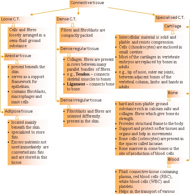
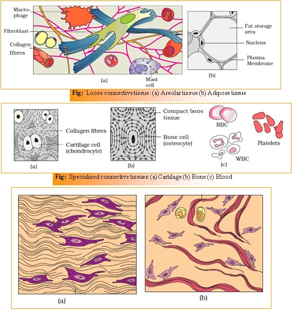
Connective Tissue
Connective tissues are most abundant and widely distributed in the body of complex animals.
They are named connective tissues because of their special function of linking and supporting other tissues/organs of the body.
In all connective tissues except blood, the cells secrete fibres of structural proteins called collagen or elastin which provide strength, elasticity and flexibility to the tissue.
These cells also secrete modified polysaccharides, which accumulate between cells and fibres and act as matrix (ground substance).
Connective tissues are classified into three types: (i) Loose connective tissue, (ii) Dense connective tissue and (iii) Specialised connective tissue.
Muscle Tissue
- Each muscle is made of many long, cylindrical fibres arranged in parallel arrays. These fibres are composed of numerous fine fibrils, called myofibrils.
- Muscle fibres contract (shorten) in response to stimulation, then relax (lengthen) and return to their uncontracted state in a coordinated fashion.
- Their action moves the body to adjust to the changes in the environment and to maintain the positions of the various parts of the body.
- In general, muscles play an active role in all the movements of the body.
- Muscles are of three types, skeletal, smooth, and cardiac.


Neural Tissue
- Neural tissue consists of neuron and neuroglial cells.
- Neural tissue exerts the greatest control over the body’s responsiveness to changing conditions.
- Neuron, an excitable cell is the unit of neural system.
- The neuroglial cells which constitute the rest of the neural system protect and support neurons.
- Neuroglia make up more than one half the volume of neural tissue in our body.
- When a neuron is suitably stimulated, an electrical disturbance is generated which swiftly travels along its plasma membrane.
- Arrival of the disturbance at the neuron’s endings, or output zone, triggers events that may cause stimulation or inhibition of adjacent neurons and other cells

ORGAN AND ORGAN SYSTEM
- Tissues organise to form organs which in turn associate to form organ systems in the multicellular organisms, this results in more efficient and coordinated system of cells.
- Each organ is made of one or more type of tissues.
- Complexity in organ and organ systems displays certain evolutionary trend.
EARTHWORM
Habits and habitat –
- Earthworm is a reddish brown terrestrial invertebrate that inhabits the upper layer of the moist soil.
- During day time, they live in burrows made by boring and swallowing the soil. In the gardens, they can be traced by their faecal deposits known as worm castings.
- The common Indian earthworms are Pheretima and
Morphology
- Long cylindrical body.
- Body is divided into many short segments which are similar (metameres about 100-120).
-
Body surfaces –
- The dorsal surface of the body is marked by a dark median mid dorsal line (dorsal blood vessel) along the longitudinal axis of the body.
- The ventral surface is distinguished by the presence of genital openings (pores).
- Anterior end consists of the mouth and the prostomium, a lobe which serves as a covering for the mouth and as a wedge to force open cracks in the soil into which the earthworm may crawl. The prostomium is sensory in function.
-
Segments and their related structures –
- The first body segment is called the peristomium (buccal segment) which contains the mouth.
- In a mature worm, 14th, 15th, 16th segments are covered by a prominent dark band of glandular tissue called clitellum. Thus the body is divisible into three prominent regions – preclitellar, clitellar and postclitellar segments.
- Four pairs of spermathecal apertures are situated on the ventro-lateral sides of the intersegmental grooves, i.e., 5th -9th
- A single female genital pore is present in the mid-ventral line of 14th
- A pair of male genital pores are present on the ventro-lateral sides of the 18th
- Numerous minute pores called nephridiopores open on the surface of the body.
- In each body segment, except the first, last and clitellum, there are rows of S-shaped setae, embedded in the epidermal pits in the middle of each segment. Setae can be extended or retracted. Their principal role is in locomotion.

Anatomy
Body Wall
- Layers are outermost thin non-cellular cuticle, epidermis, two muscle layers (circular and longitudinal) and an innermost coelomic epithelium
- The epidermis is made up of a single layer of columnar epithelial cells which contain secretory gland cells.
Alimentary canal
- It is a straight tube and runs between first to last segment of the body.
- It consists of a terminal mouth, buccal cavity (1-3 segments), muscular pharynx, oesophagus (5-7 segments), muscular gizzard (8-9 segments), stomach (9-14 segments), intestine (15th to last segment), anus.
- Gizzard helps in grinding the soil particles and decaying leaves.
- Calciferous glands, present in the stomach, neutralise the humic acid present in humus.
- A pair of short and conical intestinal caecae project from the intestine on the 26th segment.
- In intestine between 26-35 segments, an internal median fold of dorsal wall called typhlosole is present. It increases the effective area of absorption in the intestine.
Circulatory system
- Pheretima exhibits a closed type of blood vascular system, consisting of blood vessels, capillaries and heart.
- Blood is confined to the heart and blood vessels. Contractions keep blood circulating in one direction. Smaller blood vessels supply the gut, nerve cord, and the body wall.
- Blood glands are present on the 4th, 5th and 6th segments. They produce blood cells and haemoglobin which is dissolved in blood plasma.
- Blood cells are phagocytic in nature.
Respiratory system
- Earthworms lack specialised breathing devices.
- Respiratory exchange occurs through moist body surface into their blood stream.
Excretory system
- The excretory organs occur as segmentally arranged coiled tubules called nephridia.
- They are of three types (similar in structure) :
- septal nephridia – Present on both the sides of intersegmental septa of segment 15 to the last that open into intestine.
- integumentary nephridia – attached to lining of the body wall of segment 3 to the last that open on the body surface
- pharyngeal nephridia – Present as three paired tufts in the 4th, 5th and 6th segments.
- Nephridia regulate the volume and composition of the body fluids. (osmotic regulation).
- A nephridium starts out as a funnel that collects excess fluid from coelomic chamber. The funnel connects with a tubular part of the nephridium which delivers the wastes through a pore to the surface in the body wall into the digestive tube.
Nervous system
- It is basically represented by ganglia arranged segmentwise on the ventral paired nerve cord.
- The nerve cord in the anterior region (3rd and 4th segments) bifurcates, laterally encircling the pharynx and joins the cerebral ganglia dorsally to form a nerve ring.
- The cerebral ganglia alongwith other nerves in the ring integrate sensory input as well as command muscular responses of the body.
Sense organs
- eyes are absent but does possess light and touch sensitive organs.
- Worms have specialised chemoreceptors (taste receptors) which react to chemical stimuli.
- These sense organs are located on the anterior part of the worm.
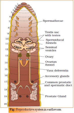
Reproductive system
- Earthworm is hermaphrodite (bisexual), i.e., testes and ovaries are present in the same individual.
- Male –
- two pairs of testes (10th, 11th segments).
- Their vasa deferentia run up to the 18th segment where they join the prostatic duct.
- Two pairs of accessory glands are present (in the 17th, 19th segments).
- The common prostrate and spermatic duct (vary differential) opens to the exterior by a pair of male genital pores on the ventro-lateral side of the 18th
- Female –
- Four pairs of spermathecae are located in 6th-9th segments (one pair in each segment). They receive and store spermatozoa during copulation.
- One pair of ovaries is attached at the inter-segmental septum of the 12th and 13th
- Ovarian funnels are present beneath the ovaries which continue into oviduct, join together and open on the ventral side as a single median female genital pore on the 14th segment.
- Fertilization –
- It is a protandrous animal with crossfertilisation.
- A mutual exchange of sperm occurs between two worms during mating. One worm has to find another worm and they mate juxtaposing opposite gonadal openings exchanging packets of sperms called spermatophores.
- Mature sperm and egg cells and nutritive fluid are deposited in cocoons produced by the gland cells of clitellum.
- Fertilisation and development occur within the cocoons which are deposited in soil.
- The ova (eggs) are fertilised by the sperm cells within the cocoon which then slips off the worm and is deposited in or on the soil.
- The cocoon holds the worm embryos.
- After about 3 weeks, each cocoon produces two to twenty baby worms with an average of four.
- Earthworms development is direct, i.e., there is no larva formed.
Economical uses –
- Earthworms are known as ‘friends of farmers’ because they make burrows in the soil and make it porous which helps in respiration and penetration of the developing plant roots. The process of increasing fertility of soil by the earthworms is called vermicomposting.
- They are also used as bait in game fishing.

COCKROACH
- Brown or black bodied animals.
- Included in class Insecta of Phylum Arthropoda.
- Bright yellow, red and green coloured cockroaches have also been reported in tropical regions.
- Size ranges from ¼ inches to 3 inches (0.6-7.6 cm) and have long antenna, legs and flat extension of the upper body wall that conceals head.
- Nocturnal, Omnivores that live in damp places throughout the world.
- They have become residents of human homes and thus are serious pests and vectors of several diseases.
Morphology
- Scientific name of the common species of cockroach, Periplaneta Americana.
- They are about 34-53 mm long with wings that extend beyond the tip of the abdomen in males.
- The body of the cockroach is segmented and divisible into three distinct regions – head, thorax and abdomen.
- The entire body is covered by a hard chitinous exoskeleton (brown in colour).
- In each segment, exoskeleton has hardened plates called sclerites (tergites dorsally and sternites ventrally) that are joined to each other by a thin and flexible articular membrane (arthrodial membrane).
-
Head –
- Head is triangular in shape and lies anteriorly at right angles to the longitudinal body axis.
- It is formed by the fusion of six segments and shows great mobility in all directions due to flexible neck.
- The head capsule bears a pair of compound eyes, a pair of thread like antennae which arise from membranous sockets lying in front of eyes. Antennae have sensory receptors that help in monitoring the environment.
- At anterior end of the head, appendages forming biting and chewing type of mouth parts are present. The mouthparts consisting of a labrum (upper lip), a pair of mandibles, a pair of maxillae and a labium (lower lip).
- A median flexible lobe, acting as tongue (hypopharynx), lies within the cavity enclosed by the mouthparts
-
Thorax –
- It consists of three parts – prothorax, mesothorax and metathorax.
- The head is connected with thorax by a short extension of the prothorax known as the neck.
- Each thoracic segment bears a pair of walking legs.
- The first pair of wings arises from mesothorax and the second pair from metathorax. Forewings (mesothoracic) called tegmina are opaque dark and leathery and cover the hind wings when at rest. The hind wings are transparent, membranous and are used in flight.
-
Abdomen –
- The abdomen in both males and females consists of 10 segments.
- In females, the 7th sternum is boat shaped and together with the 8th and 9th sterna form a brood or genital pouch whose anterior part contains female gonopore, spermathecal pores and collateral glands.
- In males, genital pouch or chamber lies at the hind end of abdomen bounded dorsally by 9th and 10th terga and ventrally by the 9th It contains dorsal anus, ventral male genital pore and gonapophysis.
- Males bear a pair of short, threadlike anal styles which are absent in females.
- In both sexes, the 10th segment bears a pair of jointed filamentous structures called anal cerci.

Anatomy
-
Digestive system –
- The alimentary canal is divided into three regions: foregut, midgut and hindgut.
- Fore gut –
- Consist of mouth, pharynx, oesophagus, crop, gizzard (Proventriculus).
- Crop is sac like structure for storing of food.
- Gizzard has an outer layer of thick circular muscles and thick inner cuticle forming six highly chitinous plate called teeth. Gizzard helps in grinding the food particles.
- The entire foregut is lined by cuticle.
- Mid gut –
- A ring of 6-8 blind tubules called hepatic or gastric caecae is present at the junction of foregut and midgut, which secrete digestive juice.
- Hind gut –
- At the junction of midgut and hindgut 100-150 yellow coloured thin filamentous Malphigian tubules are present. They help in removal of excretory products from haemolymph.
- The hindgut is broader than midgut and is differentiated into ileum, colon and rectum.
- The rectum opens out through anus.
- Blood vascular system –
- Open type circulatory system.
- Blood vessels are poorly developed and open into space (haemocoel).
- Visceral organs located in the haemocoel are bathed in blood (haemolymph).
- The haemolymph is composed of colourless plasma and haemocytes.
- Heart of cockroach consists of elongated muscular tube lying along mid dorsal line of thorax and abdomen.
- It is differentiated into funnel shaped chambers with ostia on either side.
- Blood from sinuses enter heart through ostia and is pumped anteriorly to sinuses again.
- Respiratory system –
- consists of a network of trachea, that open through 10 pairs of small holes called spiracles present on the lateral side of the body.
- Thin branching tubes (tracheal tubes subdivided into tracheoles) carry oxygen from the air to all the parts.
- The opening of the spiracles is regulated by the sphincters.
- Exchange of gases take place at the tracheoles by diffusion.
- Excretory system –
- Excretion is performed by Malpighian tubules.
- Each tubule is lined by glandular and ciliated cells.
- They absorb nitrogenous waste products and convert them into uric acid which is excreted out through the hindgut. Therefore, this insect is called uricotelic.
- In addition, the fat body, nephrocytes and urecose glands also help in excretion.
- Nervous system –
- It consists of a series of fused, segmentally arranged ganglia joined by paired longitudinal connectives on the ventral side. Three ganglia lie in the thorax, and six in the abdomen.
- The nervous system of cockroach is spread throughout the body.
- The head holds a bit of a nervous system while the rest is situated along the ventral (belly-side) part of its body. So, if the head of a cockroach is cut off, it will still live for as long as one week.
- In the head region, the brain is represented by supra-oesophageal ganglion which supplies nerves to antennae and compound eyes.
- Sense organs –
- In cockroach, the sense organs are antennae, eyes, maxillary palps, labial palps, anal cerci, etc.
- The compound eyes are situated at the dorsal surface of the head. Each eye consists of about 2000 hexagonal ommatidia. With the help of several ommatidia, a cockroach can receive several images of an object. This kind of vision is known as mosaic vision with more sensitivity but less resolution, being common during night (hence called nocturnal vision).
- Reproductive system –
- Cockroaches are dioecious and both sexes have well developed reproductive organs.
- Male reproductive system –
- It consists of a pair of testes (in the 4th -6th abdominal segments), vas deferens, ejaculatory duct, seminal vesicle.
- The ejaculatory duct opens into male gonopore situated ventral to anus.
- A characteristic mushroom shaped gland is present in the 6th-7th abdominal segments which functions as an accessory reproductive gland.
- The external genitalia are represented by male gonapophysis or phallomere (chitinous asymmetrical structures, surrounding the male gonopore).
- The sperms are stored in the seminal vesicles and are glued together in the form of bundles called spermatophores which are discharged during copulation.
- Female reproductive system –
- It consists of two large ovaries (2nd – 6th abdominal segments), oviducts, vagina, genital chamber, spermathecal.
- Each ovary is formed of a group of eight ovarian tubules or ovarioles, containing a chain of developing ova.
- A pair of spermatheca is present in the 6th segment which opens into the genital chamber.
- Sperms are transferred through spermatophores.
- Fertilization and development –
- Fertilization internal.
- Fertilized eggs are encased in capsules called oothecae. Ootheca is a dark reddish to blackish brown capsule, about 3/8″ (8 mm) long.
- They are dropped or glued to a suitable surface, usually in a crack or crevice of high relative humidity near a food source.
- On an average, females produce 9-10 oothecae, each containing 14-16 eggs.
- The development of americana is paurometabolous, meaning there is development through nymphal stage. The nymphs look very much like adults. The nymph grows by moulting about 13 times to reach the adult form.
- The next to last nymphal stage has wing pads but only adult cockroaches have wings.

FROGS
Habits and habitat
- Frogs can live both on land and in freshwater and belong to class Amphibia of phylum Chordata.
- Most common species of frog found in India is Rana tigrina.
- They do not have constant body temperature i.e.; their body temperature varies with the temperature of the environment. Such animals are called cold blooded or poikilotherms.
- They have the ability to change the colour to hide them from their enemies (camouflage). This protective coloration is called mimicry.
- They take shelter in deep burrows to protect them from extreme heat and cold. This is called as summer sleep (aestivation) and winter sleep (hibernation).
Morphology
- The skin is smooth and slippery due to the presence of mucus. The skin is always maintained in a moist condition.
- The colour of dorsal side of body is generally olive green with dark irregular spots. On the ventral side the skin is uniformly pale yellow.
- The frog never drinks water but absorb it through the skin.
- Body of a frog is divisible into head and trunk. A neck and tail are absent.
- Above the mouth, a pair of nostrils is present.
- Eyes are bulged and covered by a nictitating membrane that protects them while in water.
- On either side of eyes, a membranous tympanum (ear) receives sound signals.
- The forelimbs and hind limbs help in swimming, walking, leaping and burrowing. The hind limbs end in five digits and they are larger and muscular than fore limbs that end in four digits.
- Feet have webbed digits that help in swimming.
- Frogs exhibit sexual dimorphism. Male frogs can be distinguished by the presence of sound producing vocal sacs and also a copulatory pad on the first digit of the fore limbs which are absent in female frogs.
Anatomy
-
Digestive System –
- It consists of alimentary canal and digestive glands.
- The alimentary canal is short because frogs are carnivores and hence the length of intestine is reduced.
- Alimentary canal consists of mouth, buccal cavity, pharynx, oesophagus, stomach, intestine, rectum and cloaca.
- Food is captured by the bilobed tongue.
- Digestion of food takes place by the action of HCl and gastric juices secreted from the walls of the stomach.
- Partially digested food called chyme is passed from stomach to the first part of the intestine, the duodenum.
- Liver secretes bile that is stored in the gall bladder.
- Pancreas produces pancreatic juice containing digestive enzymes.
- The duodenum receives bile from gall bladder and pancreatic juices from the pancreas through a common bile duct.
- Bile emulsifies fat and pancreatic juices digest carbohydrates and proteins.
- Final digestion takes place in the intestine.
- Digested food is absorbed by the numerous finger-like folds in the inner wall of intestine called villi and microvilli.
- The undigested solid waste moves into the rectum and passes out through cloaca.
-
Respiratory system –
- Frogs respire on land and in the water by two different methods.
- In water, skin acts as aquatic respiratory organ (cutaneous respiration). Dissolved oxygen in the water is exchanged through the skin by diffusion.
- On land, the buccal cavity, skin and lungs act as the respiratory organs.
- The respiration by lungs is called pulmonary respiration. The lungs are a pair of elongated, pink coloured sac-like structures present in the upper part of the trunk region (thorax). Air enters through the nostrils into the buccal cavity and then to lungs.
- During aestivation and hibernation gaseous exchange takes place through skin.
-
Circulatory system –
- The vascular system of frog is well-developed closed type.
- Frogs have a lymphatic system also.
- The blood vascular system involves heart, blood vessels and blood.
- The lymphatic system consists of lymph, lymph channels and lymph nodes.
- Heart is a muscular structure situated in the upper part of the body cavity.
- It has three chambers, two atria and one ventricle and is covered by a membrane called pericardium.
- A triangular structure called sinus venosus joins the right atrium. It receives blood through the major veins called vena cava.
- The ventricle opens into a saclike conus arteriosus on the ventral side of the heart.
- The blood from the heart is carried to all parts of the body by the arteries (arterial system).
- The veins collect blood from different parts of body to the heart and form the venous system.
- Special venous connection between liver and intestine as well as the kidney and lower parts of the body are present in frogs. The former is called hepatic portal system and the latter is called renal portal system.
- The blood is composed of plasma and cells.
- The blood cells are RBC (red blood cells) or erythrocytes, WBC (white blood cells) or leucocytes and platelets.
- RBC’s are nucleated and contain red coloured pigment namely haemoglobin.
- The lymph is different from blood.
- It lacks few proteins and RBCs.
- The blood carries nutrients, gases and water to the respective sites during the circulation.
- The circulation of blood is achieved by the pumping action of the muscular heart.
-
Excretory system –
- The elimination of nitrogenous wastes is carried out by a well-developed excretory system.
- The excretory system consists of a pair of kidneys, ureters, cloaca and urinary bladder.
- Kidneys are compact, dark red and bean like structures situated a little posteriorly in the body cavity on both sides of vertebral column.
- Each kidney is composed of several structural and functional units called uriniferous tubules or nephrons.
- Two ureters emerge from the kidneys in the male frogs. The ureters act as urinogenital duct which opens into the cloaca.
- In females the ureters and oviduct open seperately in the cloaca.
- The thin-walled urinary bladder is present ventral to the rectum which also opens in the cloaca.
- The frog excretes urea and thus is a ureotelic
- Excretory wastes are carried by blood into the kidney where it is separated and excreted.
-
Endocrine system-
- The chemical coordination of various organs of the body is achieved by hormones which are secreted by the endocrine glands.
- The prominent endocrine glands found in frog are pituitary, thyroid, parathyroid, thymus, pineal body, pancreatic islets, adrenals and gonads.
-
Nervous system –
- The nervous system is organised into a central nervous system (brain and spinal cord), a peripheral nervous system (cranial and spinal nerves) and an autonomic nervous system (sympathetic and parasympathetic).
- There are ten pairs of cranial nerves arising from the brain.
- Brain is enclosed in a bony structure called brain box (cranium).
- The brain is divided into fore-brain, mid-brain and hind-brain.
- Forebrain includes olfactory lobes, paired cerebral hemispheres and unpaired diencephalon.
- The midbrain is characterised by a pair of optic lobes.
- Hind-brain consists of cerebellum and medulla oblongata.
- The medulla oblongata passes out through the foramen magnum and continues into spinal cord, which is enclosed in the vertebral column.
-
Sense organs –
- Frog has different types of sense organs, namely organs of touch (sensory papillae), taste (taste buds), smell (nasal epithelium), vision (eyes) and hearing (tympanum with internal ears).
- Eyes and internal ears are well-organised structures and the rest are cellular aggregations around nerve endings.
- Eyes in a frog are a pair of spherical structures situated in the orbit in skull. These are simple eyes (possessing only one unit).
- External ear is absent in frogs and only tympanum can be seen externally. The ear is an organ of hearing as well as balancing (equilibrium).
-
Reproductive system –
- Frogs have well organised male and female reproductive systems.
- Male reproductive system –
- It consists of a pair of yellowish ovoid testes, which are found adhered to the upper part of kidneys by a double fold of peritoneum called mesorchium.
- Vasa efferentia are 10-12 in number that arise from testes.
- They enter the kidneys on their side and open into Bidder’s canal.
- Finally, it communicates with the urinogenital duct that comes out of the kidneys and opens into the cloaca.
- The cloaca is a small, median chamber that is used to pass faecal matter, urine and sperms to the exterior.
- Female reproductive system –
- It includes a pair of ovaries. The ovaries are situated near kidneys and there is no functional connection with kidneys.
- A pair of oviduct arising from the ovaries opens into the cloaca separately.
- A mature female can lay 2500 to 3000 ova at a time.
- Fertilisation and development –
- Fertilization is external and takes place in water.
- Development involves a larval stage called tadpole.
- Tadpole undergoes metamorphosis to form the adult.
Economic importance –
- Frogs are beneficial for mankind because they eat insects and protect the crop.
- Frogs maintain ecological balance because these serve as an important link of food chain and food web in the ecosystem.
- In some countries the muscular legs of frog are used as food by man.


printable pdf file of notes is available.. for download please click on following link
CHAPTER 7: STRUCTURAL ORGANISATION IN ANIMALS
