CHAPTER 13 – PHOTOSYNTHESIS IN HIGHER PLANTS

CHAPTER 13

PHOTOSYNTHESIS IN HIGHER PLANTS

  • Green plants carry out ‘photosynthesis’, a physico-chemical process by which they use light energy to drive the synthesis of organic compounds.
  • Ultimately, all living forms on earth depend on sunlight for energy.
  • The use of energy from sunlight by plants doing photosynthesis is the basis of life on earth. Photosynthesis is important due to two reasons:

(a) it is the primary source of all food on earth.

(b) It is also responsible for the release of oxygen into the atmosphere by green plants.

  • chlorophyll (green pigment of the leaf), light and CO2 are required for photosynthesis to occur.


  • Two leaves experiment for importance of chlorophyll for starch formation – a variegated leaf or a leaf that was partially covered with black paper, and one that was exposed to light. On testing these leaves for starch it was clear that photosynthesis occurred only in the green parts of the leaves in the presence of light.
  • Half-leaf experiment for importance of CO2 for starch formation – a part of a leaf is enclosed in a test tube containing some KOH soaked cotton (which absorbs CO2), while the other half is exposed to air. The setup is then placed in light for some time. On testing for starch later in the two halves of the leaf the exposed part of the leaf tested positive for starch while the portion that was in the tube, tested negative.


Early Experiments

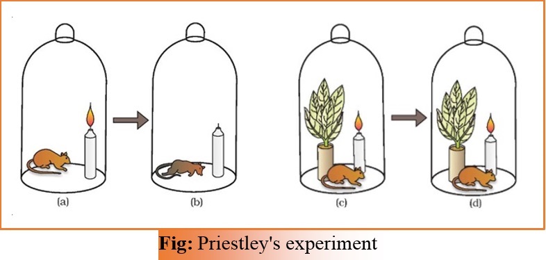
Joseph Priestley (1770)               – revealed the essential role of air in the growth of green plants

– discovered oxygen in 1774.

– Bell jar experiment

– hypothesised that Plants restore to the air whatever breathing animals and burning candles remove.

1


Jan Ingenhousz (1730-1799)      – showed that sunlight is essential to the photosynthesis.

Julius von Sachs (1854)              – provided evidence for production of glucose when plants grow.

                                                      – Glucose is usually stored as starch.

– showed that the green substance in plants is located in special bodies (later called chloroplasts) within plant cells.

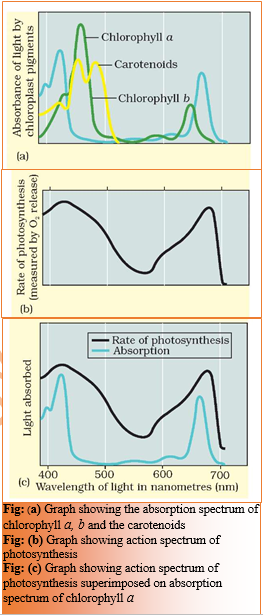
T.W Engelmann (1843 – 1909)   – Used a prism to split light into its spectral components and then illuminated a green alga, Cladophora, placed in a suspension of aerobic bacteria. The bacteria were used to detect the sites of O2 evolution.

observed that the bacteria accumulated mainly in the region of blue and red light of the split spectrum.

described first action spectrum of photosynthesis, which resembles roughly the absorption spectra of chlorophyll a and b.

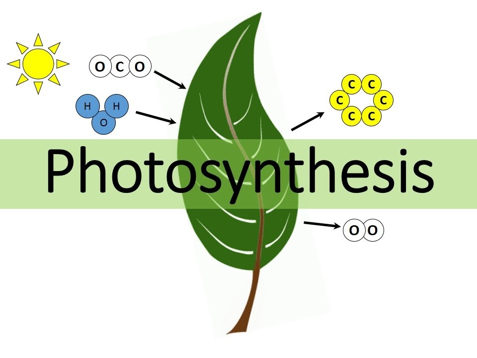
The empirical equation representing the total process of photosynthesis for oxygen evolving organisms was then understood as:

CO2 + H2O —Light –> [CH2O] + O2

where [CH2O] represented a carbohydrate (e.g., glucose, a six-carbon sugar).

Cornelius van Niel (1897-1985)  – a microbiologist

– studies of purple and green bacteria,

– demonstrated that photosynthesis is essentially a light-dependent   reaction in which hydrogen from a suitable oxidisable compound reduces carbon dioxide to carbohydrates.

2H2A + CO2Light –> 2A + CH2O + H2O

– In green plants H2O is the hydrogen donor and is oxidised to O2.

– When H2S is the hydrogen donor for purple and green sulphur bacteria, the ‘oxidation’ product is sulphur or sulphate and not O2.

– Hence, he inferred that the O2 evolved by the green plant comes from H2O, not from carbon dioxide.

– This was later proved by using radioisotopic techniques.

The correct equation, that would represent the overall process of photosynthesis is therefore:

6CO2 +12H2O —Light –> C6H12O6 + 6H2O + 6O2


SITE OF PHOTOSYNTHESIS

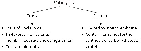
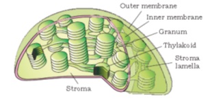
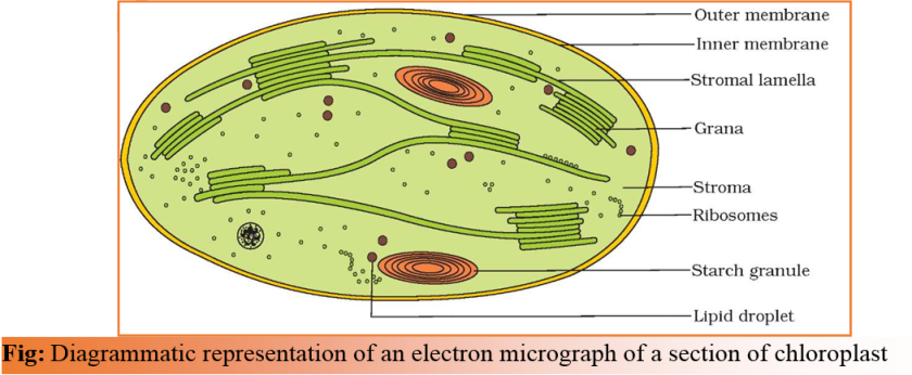
  • Photosynthesis occurs in green leaf in the chloroplasts.
  • mesophyll cells in the leaves, have a large number of chloroplasts. Usually the chloroplasts align themselves along the walls of the mesophyll cells, such that they get the optimum quantity of the incident light.
  • Within the chloroplast there is the membranous system consisting of grana, the stroma lamellae, and the fluid stroma. There is a clear division of labour within the chloroplast. The membrane system is responsible for trapping the light energy and also for the synthesis of ATP and NADPH. In stroma, enzymatic reactions incorporate CO2 into the plant leading to the synthesis of sugar, which in turn forms starch.
  • The former set of reactions, since they are directly light driven are called light reactions. The latter are not directly light driven but are dependent on the products of light reactions (ATP and NADPH). Hence, to distinguish the latter they are called, by convention, as dark reactions. However, this should not be construed to mean that they occur in darkness or that they are not light- dependent.

Screenshot (42)  HOW MANY PIGMENTS ARE INVOLVED IN PHOTOSYNTHESIS?

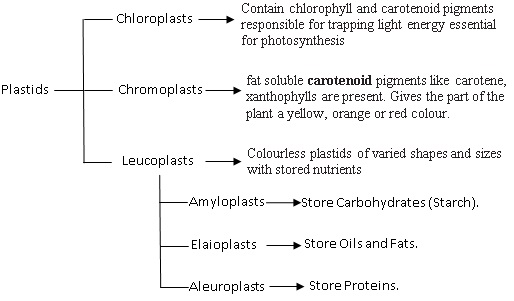
  • A chromatographic separation of the leaf pigments shows that the colour that we see in leaves is not due to a single pigment but due to four pigments:

Chlorophyll a (bright or blue green in the chromatogram),

chlorophyll b (yellow green),

xanthophylls (yellow) and

carotenoids (yellow to yellow-orange).

  • Pigments are substances that have an ability to absorb light, at specific wavelengths.
  • wavelengths at which there is maximum absorption by chlorophyll a, is in the blue and the red regions, also shows higher rate of photosynthesis. Hence, we can conclude that chlorophyll a is the chief pigment associated with photosynthesis.
  • These graphs, together, show that most of the photosynthesis takes place in the blue and red regions of the spectrum; some photosynthesis does take place at the other wavelengths of the visible spectrum.
  • Though chlorophyll is the major pigment responsible for trapping light, other thylakoid pigments like chlorophyll b, xanthophylls and carotenoids, which are called accessory pigments, also absorb light and transfer the energy to chlorophyll a. Indeed, they not only enable a wider range of wavelength of incoming light to be utilised for photosyntesis but also protect chlorophyll a from photo-oxidation.

Screenshot (43)

WHAT IS LIGHT REACTION?

  • Light reactions or the ‘Photochemical’ phase include light absorption, water splitting, oxygen release, and the formation of high-energy chemical intermediates, ATP and NADPH.
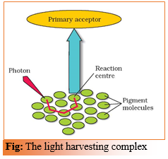
  • several complexes are involved in the process.
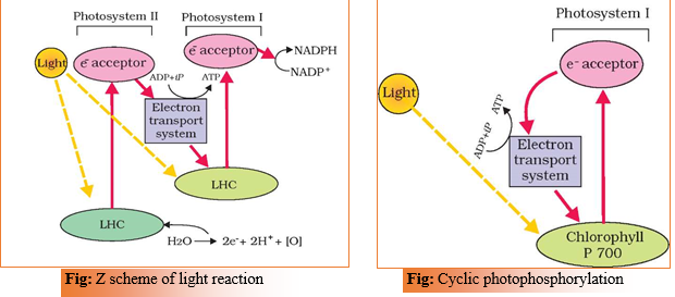
  • The pigments are organised into two discrete photochemical light harvesting complexes (LHC) within the Photosystem I (PS I) and Photosystem II (PS II). These are named in the sequence of their discovery, and not in the sequence in which they function during the light reaction.
  • The LHC are made up of hundreds of pigment molecules bound to proteins.
  • Each photosystem has all the pigments (except one molecule of chlorophyll a) forming a light harvesting system also called antennae.
  • These pigments help to make photosynthesis more efficient by absorbing different wavelengths of light. The single chlorophyll a molecule forms the reaction centre.
  • The reaction centre is different in both the photosystems.
  • In PS I the reaction centre chlorophyll a has an absorption peak at 700 nm, hence is called P700, while in PS II it has absorption maxima at 680 nm, and is called P680

Screenshot (44) THE ELECTRON TRANSPORT

  • In photosystem II the reaction centre chlorophyll a absorbs 680 nm wavelength of red light causing electrons to become excited and jump into an orbit farther from the atomic nucleus. These electrons are picked up by an electron acceptor which passes them to an electrons transport system consisting of cytochromes.
  • This movement of electrons is downhill, in terms of an oxidation-reduction or redox potential scale.
  • The electrons are not used up as they pass through the electron transport chain, but are passed on to the pigments of photosystem PS I.
  • Simultaneously, electrons in the reaction centre of PS I are also excited when they receive red light of wavelength 700 nm and are transferred to another accepter molecule that has a greater redox potential.
  • These electrons then are moved downhill again, this time to a molecule of energy-rich NADP+.
  • The addition of these electrons reduces NADP+ to NADPH + H+.
  • This whole scheme of transfer of electrons, starting from the PS II, uphill to the accepter, down the electron transport chain to PS I, excitation of electrons,transfer to another accepter, and finally down hill to NADP+ causing it to be reduced to NADPH + H+ is called the Z scheme, due to its characterstic shape. This shape is formed when all the carriers are placed in a sequence on a redox potential scale.

Screenshot (45)  Splitting of Water

  • The electrons that were moved from photosystem II must be replaced.
  • This is achieved by electrons available due to splitting of water.
  • The splitting of water is associated with the PS II; water is split into H+, [O] and electrons.
  • This creates oxygen, one of the net products of photosynthesis.
  • The electrons needed to replace those removed from photosystem I are provided by photosystem II.
  • 2H2O ——-> 4H+ + O2 + 4e
  • water splitting complex is associated with the PS II, which itself is physically located on the inner side of the membrane of the thylakoid.

Cyclic and Non-cyclic Photo-phosphorylation

  • Living organisms have the capability of extracting energy from oxidisable substances and store this in the form of bond energy.
  • Special substances like ATP, carry this energy in their chemical bonds.
  • The process of whichATP is synthesised by cells (in mitochondria and chloroplasts) is named phosphorylation.
  • Photo- phosphorylation is the synthesis of ATP from ADP and inorganic phosphate in the presence of light.
  • When the two photosystems work in a series, first PS II and then the PS I, a process called non-cyclic photo-phosphorylation occurs.
  • The two photosystems are connected through an electron transport chain, as seen earlier – in the Z scheme.
  • Both ATP and NADPH + H+ are synthesised by this kind of electron flow.
  • When only PS I is functional, the electron is circulated within the photosystem and the phosphorylation occurs due to cyclic flow of electrons.
  • A possible location where this could be happening is in the stroma lamellae.
  • While the membrane or lamellae of the grana have both PS I and PS II the stroma lamellae membranes lack PS II as well as NADP reductase enzyme.
  • The excited electron does not pass on to NADP+ but is cycled back to the PS I complex through the electron transport chain. The cyclic flow hence, results only in the synthesis of ATP, but not of NADPH + H+.
  • Cyclic photophosphorylation also occurs when only light of wavelengths beyond 680 nm are available for excitation.

Chemiosmotic Hypothesis

  • The chemiosmotic hypothesis has been put forward to explain the mechanism of synthesis of ATP.
  • Like in respiration, in photosynthesis too, ATP synthesis is linked to development of a proton gradient across a membrane.
  • This time these are membranes of the thylakoid.
  • There is one difference though, here the proton accumulation is towards the inside of the membrane, i.e., in the lumen.
  • In respiration, protons accumulate in the intermembrane space of the mitochondria when electrons move through the ETS.
  • Processes that take place during the activation of electrons and their transport to determine the steps that cause a proton gradient to develop are –

(a) Since splitting of the water molecule takes place on the inner side of the membrane, the protons or hydrogen ions that are produced by the splitting of water accumulate within the lumen of the thylakoids.

(b) As electrons move through the photosystems, protons are transported across the membrane. This happens because the primary accepter of electron which is located towards the outer side of the membrane transfers its electron not to an electron carrier but to an H carrier. Hence, this molecule removes a proton from the stroma while transporting an electron. When this molecule passes on its electron to the electron carrier on the inner side of the membrane, the proton is released into the inner side or the lumen side of the membrane.

(c) The NADP reductase enzyme is located on the stroma side of the membrane. Along with electrons that come from the accepter of electrons of PS I, protons are necessary for the reduction of NADP+ to NADPH+ H+. These protons are also removed from the stroma.

  • Hence, within the chloroplast, protons in the stroma decrease in number, while in the lumen there is accumulation of protons.
  • This creates a proton gradient across the thylakoid membrane as well as a measurable decrease in pH in the lumen.
  • This proton gradient is important because it is the breakdown of this gradient that leads to release of energy.
  • The gradient is broken down due to the movement of protons across the membrane to the stroma through the transmembrane channel of the F0 of the ATPase.
  • The ATPase enzyme consists of two parts:

one called the F0 is embedded in the membrane and forms a transmembrane channel that carries out facilitated diffusion of protons across the membrane.

The other portion is called F1 and protrudes on the outer surface of the thylakoid membrane on the side that faces the stroma. The breakdown of the gradient provides enough energy to cause a conformational change in the F1 particle of the ATPase, which makes the enzyme synthesise several molecules of energy-packed ATP.

  • Chemiosmosis requires a membrane, a proton pump, a proton gradient and ATPase.
  • Energy is used to pump protons across a membrane, to create a gradient or a high concentration of protons within the thylakoid lumen.
  • ATPase has a channel that allows diffusion of protons back across the membrane; this releases enough energy to activate ATPase enzyme that catalyses the formation of ATP.
  • Along with the NADPH produced by the movement of electrons, the ATP will be used immediately in the biosynthetic reaction taking place in the stroma, responsible for fixing CO2, and synthesis of sugars.

Screenshot (46)

WHERE ARE THE ATP AND NADPH USED?

  • The products of light reaction are ATP, NADPH and O2.
  • Of these O2 diffuses out of the chloroplast while ATP and NADPH are used to drive the processes leading to the synthesis of food, more accurately, sugars.
  • This is the biosynthetic phase of photosynthesis.
  • This process does not directly depend on the presence of light but is dependent on the products of the light reaction, i.e., ATP and NADPH, besides CO2 and H2
  • Immediately after light becomes unavailable, the biosynthetic process continues for some time, and then stops. If then, light is made available, the synthesis starts again.
  • CO2 is combined with H2O to produce (CH2O)n or sugars.
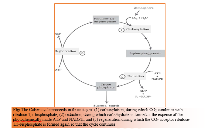
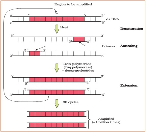
  • Melvin Calvin used radioactive 14C in algal photosynthesis studies which led to the discovery that the first CO2 fixation product was a 3-carbon organic acid or 3-phosphoglyceric acid (PGA).
  • He also contributed to working out the complete biosynthetic pathway; hence it was called Calvin cycle after him.
  • In another group of plants, first stable product of CO2 fixation is 4 carbon organic acid oxaloacetic acid or OAA.
  • The Primary Acceptor of CO2 is a 5-carbon ketose sugar – ribulose bisphosphate (RuBP).

THE CALVIN CYCLE

  • Calvin and his co-workers then worked out the whole pathway and showed that the pathway operated in a cyclic manner; the RuBP was regenerated.
  • The Calvin pathway occurs in all photosynthetic plants; it does not matter whether they have C3 or C4 (or any other) pathways.
  • The Calvin cycle can be described under three stages: carboxylation, reduction and regeneration.
  1. Carboxylation –

Carboxylation is the fixation of CO2 into a stable organic intermediate.

Carboxylation is the most crucial step of the Calvin cycle where CO2 is utilised for the carboxylation of RuBP.

This reaction is catalysed by the enzyme RuBP carboxylase which results in the formation of two molecules of 3-PGA.

Since this enzyme also has an oxygenation activity it would be more correct to call it RuBP carboxylase-oxygenase or RuBisCO.

  1. Reduction –

These are a series of reactions that lead to the formation of glucose.

The steps involve utilisation of 2 molecules of ATP for phosphorylation and two of NADPH for reduction per CO2 molecule fixed.

The fixation of six molecules of CO2 and 6 turns of the cycle are required for the removal of one molecule of glucose from the pathway.

  1. Regeneration –

Regeneration of the CO2 acceptor molecule RuBP is crucial if the cycle is to continue uninterrupted.

The regeneration steps require one ATP for phosphorylation to form RuBP.

  • Hence for every CO2 molecule entering the Calvin cycle, 3 molecules of ATP and 2 of NADPH are required.
  • It is probably to meet this difference in number of ATP and NADPH used in the dark reaction that the cyclic phosphorylation takes place.
  • To make one molecule of glucose 6 turns of the cycle are required.
  • Total 18 ATP, 12 NADPH, 12 CO2 are used synthesis of for 1 molecule of glucose.
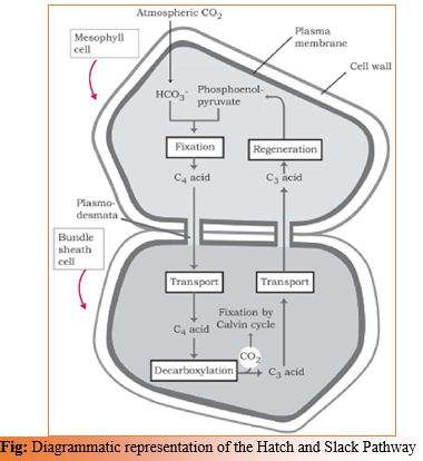
 Screenshot (47)

THE C4 PATHWAY

  • Plants that are adapted to dry tropical regions have the C4 pathway
  • Though these plants have the C4 oxaloacetic acid as the first CO2 fixation product they use the C3 pathway or the Calvin cycle as the main biosynthetic pathway.
  • C4 plants are special: They have a special type of leaf anatomy, they tolerate higher temperatures, they show a response to highlight intensities, they lack a process called photorespiration and have greater productivity of biomass.
  • The particularly large cells around the vascular bundles of the C4 pathway plants are called bundle sheath cells, and the leaves which have such anatomy are said to have ‘Kranz’ anatomy.
  • ‘Kranz’ means ‘wreath’ and is a reflection of the arrangement of cells.
  • The bundle sheath cells may form several layers around the vascular bundles; they are characterised by having a large number of chloroplasts, thick walls impervious to gaseous exchange and no intercellular spaces.
  • This pathway also known as Hatch and Slack Pathway, is a cyclic process.
  • The primary CO2 acceptor is a 3-carbon molecule phosphoenol pyruvate (PEP) and is present in the mesophyll cells. The enzyme responsible for this fixation is PEP carboxylase or PEPcase.
  • The mesophyll cells lack RuBisCO enzyme.
  • The C4 acid OAA is formed in the mesophyll cells.
  • It then forms other 4-carbon compounds like malic acid or aspartic acid in the mesophyll cells itself, which are transported to the bundle sheath cells.
  • In the bundle sheath cells these C4 acids are broken down to release CO2 and a 3-carbon molecule.
  • The 3-carbon molecule is transported back to the mesophyll where it is converted to PEP again, thus, completing the cycle.
  • The CO2 released in the bundle sheath cells enters the C3 or the Calvin pathway, a pathway common to all plants. The bundle sheath cells are rich in an enzyme Ribulose bisphosphate carboxylase-oxygenase (RuBisCO), but lack PEPcase.
  • Thus, the basic pathway that results in the formation of the sugars, the Calvin pathway, is common to the C3 and C4
  • the Calvin pathway occurs in all the mesophyll cells of the C3 plants while in the C4 plants it does not take place in the mesophyll cells but does so only in the bundle sheath cells.

Screenshot (48)

PHOTORESPIRATION

  • RuBisCO is the most abundant enzyme in the world.
  • It is characterised by the fact that its active site can bind to both CO2 and O2 – hence the name.
  • RuBisCO has a much greater affinity for CO2 than for O2.
  • This binding is competitive. It is the relative concentration of O2 and CO2 that determines which of the two will bind to the enzyme.
  • In C3 plants some O2 does bind to RuBisCO, and hence CO2 fixation is decreased. Here the RuBP instead of being converted to 2 molecules of PGA binds with O2 to form one molecule and phosphoglycolate in a pathway called photorespiration.
  • In the photorespiratory pathway, there is neither synthesis of sugars, nor of ATP. Rather it results in the release of CO2 with the utilisation of ATP.
  • In the photorespiratory pathway there is no synthesis of ATP or NADPH. Therefore, photorespiration is a wasteful process.
  • In C4 plants photorespiration does not occur. This is because they have a mechanism that increases the concentration of CO2 at the enzyme site.
  • This takes place when the C4 acid from the mesophyll is broken down in the bundle cells to release CO2 – this results in increasing the intracellular concentration of CO2.
  • In turn, this ensures that the RuBisCO functions as a carboxylase minimising the oxygenase activity.
  • Because of absence of photorespiration in C4 plants, productivity and yields are better in these plants.
  • These plants show tolerance to higher temperatures.
Characteristics C3 Plants C4 Plants
Cell type in which the Calvin cycle takes place Both Bundle sheath
Cell type in which the initial carboxylation reaction occurs Both Mesophyll
How many cell types does the leaf have that fix Co2. Three: Bundle sheath, palisade, spongy mesophyll Two: Bundle sheath and

Mesophyll

Which is the primary Co2 acceptor RuBP PEP
Number of carbons in the primary Co2 acceptor 5 3
Which is the primary Co2 fixation product PGA OAA
No. of carbons in the primary Co2 fixation product 3 4
Does the plant have RuBisCo? Yes Yes
Does the plant have PEP Case? No Yes
Which cells in the plant have Rubisco? Mesophyll/Bundle sheath/none Mesophyll/Bundle sheath/none
Co2 fixation rate under high light conditions Medium High
Whether photorespiration is present at low light intensities Sometimes Negligible
Whether photorespiration is present at high light intensities Sometimes Negligible
Whether photorespiration would be present at low CO2 concentrations High Negligible
Whether photorespiration would be present at high CO2 concentrations Sometimes Negligible
Temperature optimum 20-25C 30-40 C

FACTORS AFFECTING PHOTOSYNTHESIS

  • The rate of photosynthesis is very important in determining the yield of plants including crop plants.
  • Photosynthesis is under the influence of several factors, both internal (plant) and external.
  • The plant factors include the number, size, age and orientation of leaves, mesophyll cells and chloroplasts, internal CO2 concentration and the amount of chlorophyll. The plant or internal factors are dependent on the genetic predisposition and the growth of the plant.
  • The external factors include the availability of sunlight, temperature, CO2 concentration and water.
  • As a plant photosynthesises, all these factors will simultaneously affect its rate. Hence, though several factors interact and simultaneously affect photosynthesis or CO2 fixation, usually one factor is the major cause or is the one that limits the rate. Hence, at any point the rate will be determined by the factor available at sub-optimal levels.
  • Blackman’s (1905) Law of Limiting Factors – If a chemical process is affected by more than one factor, then its rate will be determined by the factor which is nearest to its minimal value: it is the factor which directly affects the process if its quantity is changed
  • For example, despite the presence of a green leaf and optimal light and CO2 conditions, the plant may not photosynthesise if the temperature is very low. This leaf, if given the optimal temperature, will start photosynthesising.
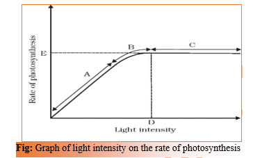
Light

  • It includes light quality, light intensity and the duration of exposure to light.
  • There is a linear relationship between incident light and CO2 fixation rates at low light intensities.
  • At higher light intensities, gradually the rate does not show further increase as other factors become limiting.
  • Light saturation occurs at 10 per cent of the full sunlight.
  • Hence, except for plants in shade or in dense forests, light is rarely a limiting factor in nature.
  • Increase in incident light beyond a point causes the breakdown of chlorophyll and a decrease in photosynthesis.

Screenshot (50)

Carbon dioxide Concentration

  • Carbon dioxide is the major limiting factor for photosynthesis.
  • The concentration of CO2 is very low in the atmosphere (between 0.03 and 0.04 per cent).
  • Increase in concentration upto 0.05 per cent can cause an increase in CO2 fixation rates; beyond this the levels can become damaging over longer periods.
  • The C3 and C4 plants respond differently to CO2 At low light conditions neither group responds to high Co2 conditions. At high light intensities, both C3 and C4 plants show increase in the rates of photosynthesis.
  • C4 plants show saturation at about 360 µlL-1 while in C3 plants saturation is seen only beyond 450 µlL-1. Thus, current availability of CO2 levels is limiting to the C3
  • The fact that C3 plants respond to higher CO2 concentration by showing increased rates of photosynthesis leading to higher productivity has been used for some greenhouse crops such as tomatoes and bell pepper. They are allowed to grow in carbon dioxide enriched atmosphere that leads to higher yields.

Temperature

  • The dark reactions being enzymatic are temperature controlled.
  • Though the light reactions are also temperature sensitive they are affected to a much lesser extent. The C4 plants respond to higher temperatures and show higher rate of photosynthesis while C3 plants have a much lower temperature optimum.
  • The temperature optimum for photosynthesis of different plants also depends on the habitat that they are adapted to.
  • Tropical plants have a higher temperature optimum than the plants adapted to temperate climates.

Water

  • Even though water is one of the reactants in the light reaction, the effect of water as a factor is more through its effect on the plant, rather than directly on photosynthesis.
  • Water stress causes the stomata to close hence reducing the CO2 Besides, water stress also makes leaves wilt, reducing the surface area of the leaves and their metabolic activity as well.

Printable PDF file is given in below link…..

CHAPTER 13 – PHOTOSYNTHESIS IN HIGHER PLANTS

CHAPTER 11 : BIOTECHNOLOGY: PRINCIPLES AND PROCESSES

Chapter 11

Biotechnology : Principles and Processes

[you can download the notes from the link given at the end of theory]

Biotechnology deals with techniques of using live organisms or enzymes from organisms to produce products and processes useful to humans.

  • Traditional form – based on natural capabilities of microorganisms. making curd, bread or wine, which are all microbe-mediated processes, could also be thought as a form of biotechnology. However, it is used in a restricted sense today,
  • Modern form – it uses genetically modified organisms to achieve the same on a larger scale. Further, many other processes/techniques are also included under biotechnology. For example, in vitro fertilisation leading to a ‘test-tube’ baby, synthesising a gene and using it, developing a DNA vaccine or correcting a defective gene, are all part of biotechnology.
  • The European Federation of Biotechnology (EFB) has given a definition of biotechnology that encompasses both traditional view and modern molecular biotechnology. The definition given by EFB is as follows:

‘The integration of natural science and organisms, cells, parts thereof, and molecular analogues for products and services’.

PRINCIPLES OF BIOTECHNOLOGY

  • Among many, the two core techniques that enabled birth of modern biotechnology are :
    • Genetic engineering: Techniques to alter the chemistry of genetic material (DNA and RNA),to introduce these into host organisms and thus change the phenotype of the host organism.
    • Maintenance of sterile (microbial contamination-free) ambience in chemical engineering processes to enable growth of only the desired microbe/eukaryotic cell in large quantities for the manufacture of biotechnological products like antibiotics, vaccines, enzymes, etc.
  • Sexual reproduction has many advantages over asexual reproduction. The former provides opportunities for variations and formulation of unique combinations of genetic setup, some of which may be beneficial to the organism as well as the population. Asexual reproduction preserves the genetic information, while sexual reproduction permits variation.
  • Traditional hybridisation procedures used in plant and animal breeding, very often lead to inclusion and multiplication of undesirable genes along with the desired genes. The techniques of genetic engineering which include creation of recombinant DNA, use of gene cloning and gene transfer, overcome this limitation and allow us to isolate and introduce only one or a set of desirable genes without introducing undesirable genes into the target organism.
  • A piece of DNA, which is somehow transferred into an alien organism, most likely would not be able to multiply itself in the progeny cells of the organism. But, when it gets integrated into the genome of the recipient, it may multiply and be inherited along with the host DNA. This is because the alien piece of DNA has become part of a chromosome, which has the ability to replicate.
  • In a chromosome there is a specific DNA sequence called the origin of replication, which is responsible for initiating replication. Therefore, for the multiplication of any alien piece of DNA in an organism it needs to be a part of a chromosome(s) which has a specific sequence known as ‘origin of replication’. Thus, an alien DNA is linked with the origin of replication, so that, this alien piece of DNA can replicate and multiply itself in the host organism. This can also be called as cloning or making multiple identical copies of any template DNA.
  • The construction of the first recombinant DNA emerged from the possibility of linking a gene encoding antibiotic resistance with a native plasmid (autonomously replicating circular extra-chromosomal DNA) of  Salmonella typhimurium.
  • Stanley Cohen and Herbert Boyer accomplished this in 1972 by isolating the antibiotic resistance gene by cutting out a piece of DNA from a plasmid which was responsible for conferring antibiotic resistance.
  • The cutting of DNA at specific locations became possible with the discovery of the so-called ‘molecular scissors’- restriction enzymes.
  • The cut piece of DNA was then linked with the plasmid DNA. These plasmid DNA act as vectors to transfer the piece of DNA attached to it. A plasmid can be used as vector to deliver an alien piece of DNA into the host organism.
  • The linking of antibiotic resistance gene with the plasmid vector became possible with the enzyme DNA ligase, which acts on cut DNA molecules and joins their ends. This makes a new combination of circular autonomously replicating DNA created in vitro and is known as recombinant DNA.
  • When this DNA is transferred into Escherichia coli, a bacterium closely related to Salmonella, it could replicate using the new host’s DNA polymerase enzyme and make multiple copies. The ability to multiply copies of antibiotic resistance gene in coli was called cloning of antibiotic resistance gene in E. coli.
  • there are three basic steps in genetically modifying an organism
    • identification of DNA with desirable genes;
    • introduction of the identified DNA into the host;
    • maintenance of introduced DNA in the host and transfer of the DNA to its progeny.

TOOLS OF RECOMBINANT DNA TECHNOLOGY

Key tools of Recombinant DNA technology are – restriction enzymes, polymerase enzymes, ligases, vectors and the host organism.

  1. Restriction Enzymes

  • In 1963, the two enzymes responsible for restricting the growth of bacteriophage in Escherichia coli were isolated. One of these added methyl groups to DNA, while the other cut DNA. The later was called restriction endonuclease.
  • The first restriction endonuclease isolated – Hind II.
  • Restriction endonuclease cut DNA molecules at a particular point by recognising a specific sequence of base pairs. This specific base sequence is known as the recognition sequence.(For Hind II – sequence of 6 base pairs).
  • Today we know more than 900 restriction enzymes that have been isolated from over 230 strains of bacteria each of which recognise different recognition sequences.

Naming of enzymes –

  • First letter of the name comes from the genes
  • The second two letters come from the species of the prokaryotic cell from which they were isolated, e.g., EcoRI comes from Escherichia coli RY 13.
  • Next letter derived from the name of strain.
  • Roman numbers following the names indicate the order in which the enzymes were isolated from that strain of bacteria.

Action of enzyme –

  • Restriction enzymes belong to a larger class of enzymes called nucleases. These are of two kinds; exonucleasesand endonucleases.
  • Exonucleases remove nucleotides from the ends of the DNA whereas, endonucleases make cuts at specific positions within the DNA.
  • Each restriction endonuclease functions by ‘inspecting’ the length of a DNA sequence. Once it finds its specific recognition sequence, it will bind to the DNA and cut each of the two strands of the double helix at specific points in their sugar -phosphate backbones.
  • Each restriction endonuclease recognises a specific palindromic nucleotide sequences in the DNA.
  • The palindrome in DNA is a sequence of base pairs that reads same on the two strands when orientation of reading is kept the same. For example, the following sequences reads the same on the two strands in 5→3 This is also true if read in the 3→5direction.

5—— GAATTC —— 3

3—— CTTAAG —— 5

  • Restriction enzymes cut the strand of DNA a little away from the centre of the palindrome sites, but between the same two bases on the opposite strands. This leaves single stranded portions at the ends. There are overhanging stretches called sticky ends on each strand.
  • These are named so because they form hydrogen bonds with their complementary cut counterparts. This stickiness of the ends facilitates the action of the enzyme DNA ligase.
  • Restriction endonucleases are used in genetic engineering to form ‘recombinant’ molecules of DNA, which are composed of DNA from different sources/genomes.
  • When cut by the same restriction enzyme, the resultant DNA fragments have the same kind of ‘sticky-ends’ and, these can be joined together (end-to-end) using DNA ligases .
  • Normally, unless one cuts the vector and the source DNA with the same restriction enzyme, the recombinant vector molecule cannot be created.

1.jpg

Fig: Steps in formation of recombinant DNA by action of restriction endonuclease enzyme – EcoRI

1.jpg

Fig: Diagrammatic representation of recombinant DNA technology


Separation and isolation of DNA fragments :

  • The cutting of DNA by restriction endonucleases results in the fragmentes of DNA. These fragments can be separated by a technique known as gel electrophoresis.
  • Since DNA fragments are negatively charged molecules they can be separated by forcing them to move towards the anode under an electric field through a medium/matrix. Nowadays the most commonly used matrix is agarose which is a natural polymer extracted from sea weeds.
  • The DNA fragments separate (resolve) according to their size through sieving effect provided by the agarose gel. Hence, the smaller the fragment size, the farther it moves.
  • The separated DNA fragments can be visualised only after staining the DNA with a compound known as ethidium bromide followed by exposure to UV radiation.
  • We can see bright orange coloured bands of DNA in aethidium bromide stained gel exposed to UV light.
  • The separated bands of DNA are cut out from the agarose gel and extracted from the gel piece. This step is known as elution. The DNA fragments purified in this way are used in constructing recombinant DNA by joining them with cloning vectors.

1.jpg

Fig: A typical agarose gel electrophoresis showing migration of undigested (lane 1) and digested set of DNA fragments (lane 2 to 4)
  1. Cloning Vectors

  • Plasmids and bacteriophages have the ability to replicate within bacterial cells independent of the control of chromosomal DNA.
  • Bacteriophages because of their high number per cell, have very high copy numbers of their genome within the bacterial cells.
  • If we are able to link an alien piece of DNA with bacteriophage or plasmid DNA, we can multiply its numbers equal to the copy number of the plasmid or bacteriophage.
  • Vectors used at present, are engineered in such way that they help easy linking of foreign DNA and selection of recombinants from non-recombinants.

Features required to facilitate cloning into a vector.

Origin of replication (ori):

  • This is a sequence from where replication starts and any piece of DNA when linked to this sequence can be made to replicate within the host cells.
  • This sequence is also responsible for controlling the copy number of the linked DNA.
  • So, if one wants to recover many copies of the target DNA it should be cloned in a vector whose origin support high copy number.

    Selectable marker :

  • In addition to ‘ori’, the vector requires a selectable marker, which helps in identifying and eliminating nontransformants and selectively permitting the growth of the transformants.
  • Transformation is a procedure through which a piece of DNA is introduced in a host bacterium.
  • Normally, the genes encoding resistance to antibiotics such as ampicillin, chloramphenicol, tetracycline or kanamycin, etc., are considered useful selectable markers for coli. The normal E. coli cells do not carry resistance against any of these antibiotics.

    Cloning sites:

  • In order to link the alien DNA, the vector needs to have very few, preferably single, recognition sites for the commonly used restriction enzymes.
  • Presence of more than one recognition sites within the vector will generate several fragments, which will complicate the gene cloning.
  • The ligation of alien DNA is carried out at a restriction site present in one of the two antibiotic resistance
  • For example, you can ligate a foreign DNA at the Bam H I site of tetracycline resistance gene in the vector pBR322. The recombinant plasmids will lose tetracycline resistance due to insertion of foreign DNA but can still be selected out from non-recombinant ones by plating the transformants on ampicillin containing medium. The transformants growing on ampicillin containing medium are then transferred on a medium containing tetracycline. The recombinants will grow in ampicillin containing medium but not on that containing tetracycline. But, nonrecombinants will grow on the medium containing both the antibiotics. In this case, one antibiotic resistance gene helps in selecting the transformants, whereas the other antibiotic resistance gene gets ‘inactivated due to insertion’ of alien DNA, and helps in selection of recombinants.
  • Selection of recombinants due to inactivation of antibiotics is a cumbersome procedure because it requires simultaneous plating on two plates having different antibiotics. Therefore, alternative selectable markers have been developed which differentiate recombinants from non-recombinants on the basis of their ability to produce colour in the presence of a chromogenic substrate.
  • In this, a recombinant DNA is inserted within the coding sequence of an enzyme, a-galactosidase. This results into inactivation of the enzyme, which is referred to as insertional inactivation. The presence of a chromogenic substrate gives blue coloured colonies if the plasmid in the bacteria does not have an insert. Presence of insert results into insertional inactivation of the a-galactosidase and the colonies do not produce any colour, these are identified as recombinant colonies.

    Vectors for cloning genes in plants and animals :

  • Viruses and bacteria are used to transfer genes into plants and animals which transform eukaryotic cells and force them to do what the bacteria or viruses want.
  • For example, Agrobacterioumtumifaciens, a pathogen of several dicot plants is able to deliver a piece of DNA known as ‘T-DNA’ to transform normal plant cells into a tumor and direct these tumor cells to produce the chemicals required by the pathogen.
  • Similarly, retroviruses in animals have the ability to transform normal cells into cancerous
  • A better understanding of the art of delivering genes by pathogens in their eukaryotic hosts has generated knowledge to transform these tools of pathogens into useful vectors for delivering genes of interest to humans.
  • The tumor inducing (Ti) plasmid of Agrobacterium tumifacienshas now been modified into a cloning vector which is no more pathogenic to the plants but is still able to use the mechanisms to deliver genes of our interest into a variety of plants. Similarly, retroviruses have also been disarmed and are now used to deliver desirable genes into animal cells. So, once a gene or a DNA fragment has been ligated into a suitable vector it is transferred into a bacterial, plant or animal host (where it multiplies).
    1.jpg
Fig: E. coli cloning vector pBR322 showing restriction sites (Hind III, EcoR I, BamH I, Sal I, PvuII, PstI, ClaI), ori and antibiotic resistance genes (ampR and tetR). Rop codes for the proteins involved in the replication of the plasmid.
  1. Competent Host (For Transformation with Recombinant DNA)

  • Since DNA is a hydrophilic molecule, it cannot pass through cell membranes. In order to force bacteria to take up the plasmid, the bacterial cells must first be made ‘competent’ to take up DNA.
  • This is done by treating them with a specific concentration of a divalent cation, such as calcium, which increases the efficiency with which DNA enters the bacterium through pores in its cell wall.
  • Recombinant DNA can then be forced into such cells by incubating the cells with recombinant DNA on ice, followed by placing them briefly at 420oC (heat shock), and then putting them back on ice. This enables the bacteria to take up the recombinant DNA.
  • In micro-injection method, recombinant DNA is directly injected into the nucleus of an animal cell.
  • In another method, suitable for plants, cells are bombarded with high velocity micro-particles of gold or tungsten coated with DNA in a method known as biolisticsor gene gun.
  • And the last method uses ‘disarmed pathogen’ vectors, which when allowed to infect the cell, transfer the recombinant DNA into the host.

PROCESSES OF RECOMBINANT DNA TECHNOLOGY

Recombinant DNA technology involves several steps in specific sequence such as –

  • isolation of DNA,
  • fragmentation of DNA by restriction endonucleases,
  • isolation of a desired DNA fragment,
  • ligation of the DNA fragment into a vector,
  • transferring the recombinant DNA into the host,
  • culturing the host cells in a medium at large scale and
  • extraction of the desired product.
  1. Isolation of the Genetic Material (DNA)

  • Nucleic acid is the genetic material of all organisms without exception. In majority of organisms this is deoxyribonucleic acid or DNA.
  • In order to cut the DNA with restriction enzymes, it needs to be in pure form, free from other macro-molecules. Since the DNA is enclosed within the membranes, we have to break the cell open to release DNA along with other macromolecules such as RNA, proteins, polysaccharides and also lipids. This can be achieved by treating the bacterial cells/plant or animal tissue with enzymes such as lysozyme (bacteria), cellulase(plant cells), chitinase(fungus).
  • genes are located on long molecules of DNA interwined with proteins such as histones.
  • RNA can be removed by treatment with ribonuclease whereas proteins can be removed by treatment with protease. Other molecules can be removed by appropriate treatments and purified DNA ultimately precipitates out after the addition of chilled ethanol. This can be seen as collection of fine threads in the suspension.

1.jpg

Fig: DNA thatseparates out can beremoved by spooling
  1. Cutting of DNA at Specific Locations

  • Restriction enzyme digestions are performed by incubating purified DNA molecules with the restriction enzyme, at the optimal conditions for that specific enzyme.
  • Agarose gel electrophoresis is employed to check the progression of a restriction enzyme digestion. DNA is a negatively charged molecule, hence it moves towards the positive electrode (anode).
  • The process is repeated with the vector DNA also.
  • The joining of DNA involves several processes. After having cut the source DNA as well as the vector DNA with a specific restriction enzyme, the cut out ‘gene of interest’ from the source DNA and the cut vector with space are mixed and ligase is added. This results in the preparation of recombinant DNA.
  1. Amplification of Gene of Interest using PCR (Polymerase Chain Reaction)

  • In this reaction, multiple copies of the gene (or DNA) of interest is synthesisedin vitro using two sets of primers (small chemically synthesised oligonucleotides that are complementary to the regions of DNA) and the enzyme DNA polymerase.
  • The enzyme extends the primers using the nucleotides provided in the reaction and the genomic DNA as template.
  • If the process of replication of DNA is repeated many times, the segment of DNA can be amplified to approximately billion times.
  • Such repeated amplification is achieved by the use of a thermostable DNA polymerase (isolated from a bacterium, Thermusaquaticus), which remain active during the high temperature induced denaturation of double stranded DNA.
  • The amplified fragment if desired can now be used to ligate with a vector for further cloning.

1.jpg

Fig: Polymerase chain reaction (PCR) : Each cycle has three steps: (i) Denaturation;

(ii) Primer annealing; and (iii) Extension of primers

  1. Insertion of Recombinant DNA into the Host Cell/Organism

  • There are several methods of introducing the ligated DNA into recipient cells. Recipient cells after making them ‘competent’ to receive, take up DNA present in its surrounding.
  • So, if a recombinant DNA bearing gene for resistance to an antibiotic (e.g., ampicillin) is transferred into coli cells, the host cells become transformed into ampicillin-resistant cells. If we spread the transformed cells on agar plates containing ampicillin, only transformants will grow, untransformed recipient cells will die. Since, due to ampicillin resistance gene, one is able to select a transformed cell in the presence of ampicillin. The ampicillin resistance gene in this case is called a selectable marker.
  1. Obtaining the Foreign Gene Product

  • When you insert a piece of alien DNA into a cloning vector and transfer it into a bacterial, plant or animal cell, the alien DNA gets multiplied.
  • In almost all recominant technologies, the ultimate aim is to produce a desirable protein. Hence, there is a need for the recombinant DNA to be expressed.
  • The foreign gene gets expressed under appropriate conditions. The expression of foreign genes in host cells involve understanding many technical details.
  • After having cloned the gene of interest and having optimised the conditions to induce the expression of the target protein, one has to consider producing it on a large scale.
  • If any protein encoding gene is expressed in a heterologous host, is called a recombinant protein.
  • The cells harbouring cloned genes of interest may be grown on a small scale in the laboratory. The cultures may be used for extracting the desired protein and then purifying it by using different separation techniques.
  • The cells can also be multiplied in a continuous culture system wherein the used medium is drained out from one side while fresh medium is added from the other to maintain the cells in their physiologically most active log/exponential phase. This type of culturing method produces a larger biomass leading to higher yields of desired protein.
  • Small volume cultures cannot yield appreciable quantities of products. To produce in large quantities, the development of bioreactors, where large volumes (100-1000 litres) of culture can be processed, was required. Thus, bioreactors can be thought of as vessels in which raw materials are biologically converted into specific products, individual enzymes, etc., using microbial plant, animal or human cells. A bioreactor provides the optimal conditions for achieving the desired product by providing optimum growth conditions (temperature, pH, substrate, salts, vitamins, oxygen).
  • A stirred-tank reactor is usually cylindrical or with a curved base to facilitate the mixing of the reactor contents. The stirrer facilitates even mixing and oxygen availability throughout the bioreactor. Alternatively air can be bubbled through the reactor.
  • The bioreactor has an agitator system, an oxygen delivery system and a foam control system, a temperature control system, pH control system and sampling ports so that small volumes of the culture can be withdrawn periodically.

1.jpg

Fig: (a) Simple stirred-tank bioreactor; (b) Sparged stirred-tank bioreactor through whichsterile air bubbles are sparged


  1. Downstream Processing
  • After completion of the biosynthetic stage, the product has to be subjected through a series of processes before it is ready for marketing as a finished The processes include separation and purification, which are collectively referred to as downstream processing.
  • The product has to be formulated with suitable preservatives. Such formulation has to undergo thorough clinical trials as in case of drugs. Strict quality control testing for each product is also required. The downstream processing and quality control testing vary from product to product.

To download notes in pdf format please click on the following link.

CHAPTER 11 : BIOTECHNOLOGY: PRINCIPLES AND PROCESSES

CHAPTER 2 : SEXUAL REPRODUCTION IN FLOWERING PLANTS

CHAPTER 2

SEXUAL REPRODUCTION IN FLOWERING PLANTS

[you can download the notes from the link given at the end of theory]

  • All flowering plants show sexual reproduction.

Flower – A Fascinating Organ of Angiosperms

  • Flowers are objects of aesthetic, ornamental, social, religious and cultural value – they have always been used as symbols for conveying important human feelings such as love, affection, happiness, grief, mourning, etc.

1

  • Pre-fertilisation: Structures and Events

  • Several hormonal and structural changes are initiated which lead to the differentiation and further development of the floral primordium.
  • Inflorescences are formed which bear the floral buds and then the flowers.
  • In the flower the male and female reproductive structures, the androecium and the gynoecium differentiate and develop.
  • The androecium consists of a whorl of stamens representing the male reproductive organ and the gynoecium represents the female reproductive organ.

Stamen, Microsporangium and Pollen Grain

  • A typical stamen has two parts –

the long and slender stalk called the filament,

and the terminal generally bilobed structure called the anther.

  • The proximal end of the filament is attached to the thalamus or the petal of the flower.
  • The number and length of stamens are variable in flowers of different species.
  • A typical angiosperm anther is bilobed with each lobe having two theca, i.e., they are
  • The anther is a four-sided (tetragonal) structure consisting of four microsporangia located at the corners, two in each lobe. tetrasporangiate
  • The microsporangia develop further and become pollen sacs. They extend longitudinally all through the length of an anther and are packed with pollen grains.

1

1.jpg

Structure of microsporangium:

  • In a transverse of an anther section, a typical microsporangium appears nearcircular in outline.
  • It is generally surrounded by four wall layers :- the epidermis, endothecium, middle layers and the tapetum.
  • The outer three wall layers perform the function of protection and help in dehiscence of anther to release the pollen.
  • The innermost wall layer is the It nourishes the developing pollen grains. Cells of the tapetum possess dense cytoplasm and generally have more than one nucleus.
  • When the anther is young, a group of compactly arranged homogenous cells called the sporogenous tissue occupies the centre of each microsporangium.

Microsporogenesis :

  • As the anther develops, the cells of the sporogenous tissue undergo meiotic divisions to form microspore tetrads, which is haploid.
  • As each cell of the sporogenous tissue is capable of giving rise to a microspore tetrad. Each one is a potential pollen or microspore mother cell (PMC).
  • The process of formation of microspores from a pollen mother cell through meiosis is called
  • The microspores, as they are formed, are arranged in a cluster of four cells-the microspore tetrad.
  • As the anthers mature and dehydrate, the microspores dissociate from each other and develop into pollen grains.
  • Inside each microsporangium several thousands of microspores or pollen grains are formed that are released with the dehiscence of anther.


Pollen grain:

  • The pollen grains represent the male gametophytes.
  • Pollen grains are generally spherical measuring about 25-50 micrometers in diameter.
  • It has a prominent two-layered wall.
    • The hard outer layer called the exine is made up of sporopollenin which is one of the most resistant organic material known. It can withstand high temperatures and strong acids and alkali. No enzyme that degrades sporopollenin is so far known. Pollen grain exine has prominent apertures called germ pores where sporopollenin is absent. Pollen grains are well- preserved as fossils because of the presence of sporopollenin. The exine exhibits a fascinating array of patterns and designs.
    • The inner wall of the pollen grain is called the It is a thin and continuous layer made up of cellulose and pectin.
  • The cytoplasm of pollen grain is surrounded by a plasma membrane.
  • When the pollen grain is mature it contains two cells, the vegetative cell and generative cell.
    • The vegetative cell is bigger, has abundant food reserve and a large irregularly shaped nucleus.
    • The generative cell is small and floats in the cytoplasm of the vegetative cell. It is spindle shaped with dense cytoplasm and a nucleus.
  • In over 60 per cent of angiosperms, pollen grains are shed at this 2-celled stage. In the remaining species, the generative cell divides mitotically to give rise to the two male gametes before pollen grains are shed (3-celled stage).

 1.jpg

1.jpg

  • Pollen grains of many species cause severe allergies and bronchial afflictions in some people often leading to chronic respiratory disorders – asthma, bronchitis, etc.
  • It may be mentioned that Parthenium or carrot grass that came into India as a contaminant with imported wheat, has become ubiquitous in occurrence and causes pollen allergy.
  • Pollen grains are rich in nutrients. It has become a fashion in recent years to use pollen tablets as food supplements.
  • In western countries, a large number of pollen products in the form of tablets and syrups are available in the market.
  • Pollen consumption has been claimed to increase the performance of athletes and race horses.
  • When once they are shed, pollen grains have to land on the stigma before they lose viability if they have to bring about fertilisation.
  • The period for which pollen grains remain viable is highly variable and to some extent depends on the prevailing temperature and humidity.
  • In some cereals such as rice and wheat, pollen grains lose viability within 30 minutes of their release, and in some members of Rosaceae, Leguminoseae and Solanaceae, they maintain viability for months.
  • It is possible to store pollen grains of a large number of species for years in liquid nitrogen (-1960C). Such stored pollen can be used as pollen banks, similar to seed banks, in crop breeding programmes.

1.jpg


The Pistil, Megasporangium (ovule) and Embryo sac (Female gametophyte):

  • The gynoecium represents the female reproductive part of the flower.
  • It may consist of a single pistil (monocarpellary) or may have more than one pistil (multicarpellary). When there are more than one, the pistils may be fused together (syncarpous) or may be free (apocarpous).
  • Each pistil has three parts – the stigma, style and ovary.

The stigma serves as a landing platform for pollen grains.

The style is the elongated slender part beneath the stigma.

The basal bulged part of the pistil is the ovary. Inside the ovary is the ovarian cavity (locule). The placenta is located inside the ovarian cavity.

  • Arising from the placenta are the megasporangia, commonly calledovules.
  • The number of ovules in an ovary may be one (wheat, paddy, mango) to many (papaya, water melon, orchids).

The Megasporangium (Ovule):

  • The ovule is a small structure attached to the placenta by means of a stalk calledfunicle.
  • The body of the ovule fuses with funicle in the region calledhilum. Thus, hilum represents the junction between ovule and funicle.
  • Each ovule has one or two protective envelopes calledinteguments. Integuments encircle the ovule except at the tip where a small opening called themicropyle is organised.
  • Opposite the micropylar end, is thechalaza, representing the basal part of the ovule.
  • Enclosed within the integuments is a mass of cells called thenucellus. Cells of the nucellus have abundant reserve food materials.
  • Located in the nucellus is theembryo sac or female gametophyte.
  • An ovule generally has a single embryo sac formed from a megaspore through reduction division.

Megasporogenesis:

  • The process of formation of megaspores from the megaspore mother cell is called
  • Ovules generally differentiate a single megaspore mother cell (MMC) in the micropylar region of the nucellus. It is a large cell containing dense cytoplasm and a prominent nucleus.
  • The MMC undergoes meiotic division.Meiosis results in the production of four megaspores.

Female gametophyte:

  • In a majority of flowering plants, one of the megaspores is functional while the other three degenerate.
  • Only the functional megaspore develops into the female gametophyte (embryo sac). This method of embryo sac formation from a single megaspore is termed monosporic
  • Ploidy of the cells of the

nucellus – 2n,

MMC – 2n,

the functional megaspore – n,

female gametophyte – n.

  • Process of development –

    The nucleus of the functional megaspore divides mitotically to form two nuclei which move to the opposite poles, forming the 2-nucleate embryo sac. Two more sequential mitotic nuclear divisions result in the formation of the 4-nucleate and later the 8-nucleate stages of the embryo sac.

these mitotic divisions are strictly free nuclear. (nuclear divisions are not followed immediately by cell wall formation.)

After the 8-nucleate stage, cell walls are laid down leading to the organisation of the typical female gametophyte or embryo sac.

  • Structure –

Six of the eight nuclei are surrounded by cell walls and organised into cells; the remaining two nuclei, called polar nuclei are situated below the egg apparatus in the large central cell.

Three cells are grouped together at the micropylar end and constitute the egg apparatus. The egg apparatus, in turn, consists of two synergids and one egg cell.

The synergids have special cellular thickenings at the micropylar tip called filiform apparatus, which play an important role in guiding the pollen tubes into the synergid.

Three cells are at the chalazal end and are called the antipodals.

The large central cell, as mentioned earlier, has two polar nuclei.

Thus, a typical angiosperm embryo sac, at maturity, though 8-nucleate is 7-celled.

1


1.jpg


Pollination

  • both male and female gametes are non-motile, so they have to be brought together for fertilisation to occur.
  • The transfer of pollen grains (shed from the anther) to the stigma of a pistil is termedpollination.
  • Kinds of Pollination : Depending on the source of pollen, pollination can be divided into three types.
    • Autogamy : Transfer of pollen grains from the anther to the stigma of the same flower. In a normal flower which opens and exposes the anthers and the stigma, complete autogamy is rather rare. Autogamy in such flowers requires synchrony in pollen release and stigma receptivity and also, the anthers and the stigma should lie close to each other so that self-pollination can occur.

Some plants such as Viola (common pansy), Oxalis, and Commelina produce two types of flowers –

chasmogamous flowers which are similar to flowers of other species with exposed anthers and stigma, and

cleistogamous flowers which do not open at all. In such flowers, the anthers and stigma lie close to each other. When anthers dehisce in the flower buds, pollen grains come in contact with the stigma to effect pollination. Thus, cleistogamous flowers are invariably autogamous as there is no chance of cross-pollen landing on the stigma. Cleistogamous flowers produce assured seed-set even in the absence of pollinators.

  • Geitonogamy – Transfer of pollen grains from the anther to the stigma of another flower of the same plant. Although geitonogamy is functionally cross-pollination involving a pollinating agent, genetically it is similar to autogamy since the pollen grains come from the same plant.
  • Xenogamy – Transfer of pollen grains from anther to the stigma of a different plant. This is the only type of pollination which during pollination brings genetically different types of pollen grains to the stigma.

1.jpg

 Agents of Pollination:

  • Plants use two abiotic (wind and water) and one biotic (animals) agents to achieve pollination.
  • Majority of plants use biotic agents for pollination. Only a small proportion of plants use abiotic agents.
  • Pollen grains coming in contact with the stigma is a chance factor in both wind and water pollination. To compensate for this uncertainties and associated loss of pollen grains, the flowers produce enormous amount of pollen when compared to the number of ovules available for pollination.
  • Wind poliination –
    • Pollination by wind is more common amongst abiotic pollinations.
    • Wind pollination requires that the pollen grains are light and non-sticky so that they can be transported in wind currents.
    • These plants often possess well-exposed stamens (so that the pollens are easily dispersed into wind currents) and large often-feathery stigma to easily trap air-borne pollen grains.
    • Wind- pollinated flowers often have a single ovule in each ovary and numerous flowers packed into an inflorescence.
    • g., corn cob, grasses.
  • Water pollination –
    • Pollination by water is quite rare in flowering plants, present in mostly monocotyledons.
    • water is a regular mode of transport for the male gametes among the lower plant groups such as algae, bryophytes and pteridophytes.
    • g., Vallisneria and Hydrilla (fresh water) and Zostera (marine sea-grasses).
    • Not all aquatic plants use water for pollination.
    • In a majority of aquatic plants such as water hyacinth and water lily, the flowers emerge above the level of water and are pollinated by insects or wind as in most of the land plants.
    • In Vallisneria, the female flower reach the surface of water by the long stalk and the male flowers or pollen grains are released on to the surface of water. They are carried passively by water currents; some of them eventually reach the female flowers and the stigma.
    • In seagrasses, female flowers remain submerged in water and the pollen grains are released inside the water. Pollen grains in many such species are long, ribbon like and they are carried passively inside the water; some of them reach the stigma and achieve pollination.
    • In most of the water-pollinated species, pollen grains are protected from wetting by a mucilaginous covering.
    • Both wind and water pollinated flowers are not very colourful and do not produce nectar.
  • Animal pollination –
    • Mode of pollination in majority of flowering plants.
    • Bees, butterflies, flies, beetles, wasps, ants, moths, birds (sunbirds and humming birds) and bats are the common pollinating agents.
    • Among the animals, insects, particularly bees are the dominant biotic pollinating agents.
    • Even larger animals such as some primates (lemurs), arboreal (tree-dwelling) rodents, or even reptiles (gecko lizard and garden lizard) are also pollinators in some species.
    • Often flowers of animal- pollinated plants are specifically adapted for a particular species of animal.
    • Majority of insect-pollinated flowers are large, colourful, fragrant and rich in nectar.
    • When the flowers are small, a number of flowers are clustered into an inflorescence to make them conspicuous.
    • Animals are attracted to flowers by colour and/or fragrance.
    • The flowers pollinated by flies and beetles secrete foul odours to attract these animals.
    • To sustain animal visits, the flowers have to provide rewards to the animals. Nectar and pollen grains are the usual floral rewards.
    • For harvesting the reward(s) from the flower the animal visitor comes in contact with the anthers and the stigma. The body of the animal gets a coating of pollen grains, which are generally sticky in animal pollinated flowers. When the animal carrying pollen on its body comes in contact with the stigma, it brings about pollination.
    • In some species floral rewards are in providing safe places to lay eggs; e.g., tallest flower of Amorphophallus (6 feet in height).
    • A similar relationship exists between a species of moth and the plant Yucca where both species – moth and the plant – cannot complete their life cycles without each other. The moth deposits its eggs in the locule of the ovary and the flower, in turn, gets pollinated by the moth. The larvae of the moth come out of the eggs as the seeds start developing.
    • Other examples of insect pollinated plants – Cucumber, Mango, PeepaL, Coriander, Papaya, Onion, Lobia, Cotton, Tobacco, Rose, Lemon, Eucalyptus, Banana.
    • Pollen/ Nectar robbers – Many insects may consume pollen or the nectar without bringing about pollination. Such floral visitors are referred to as pollen/nectar robbers.

Outbreeding Devices:

  • Majority of flowering plants produce hermaphrodite flowers and pollen grains are likely to come in contact with the stigma of the same flower.
  • Continued self-pollination result in inbreeding depression.
  • Flowering plants have developed many devices to discourage self- pollination and to encourage cross-pollination.
  • In some species, pollen release and stigma receptivity are not synchronised. Either the pollen is released before the stigma becomes receptive or stigma becomes receptive much before the release of pollen.
  • In some other species, the anther and stigma are placed at different positions so that the pollen cannot come in contact with the stigma of the same flower. Both these devices prevent autogamy.
  • self-incompatibility – This is a genetic mechanism and prevents self-pollen (from the same flower or other flowers of the same plant) from fertilising the ovules by inhibiting pollen germination or pollen tube growth in the pistil.
  • production of unisexual flowers.

If both male and female flowers are present on the same plant such as castor and maize (monoecious), it prevents autogamy but not geitonogamy.

In several species such as papaya, male and female flowers are present on different plants, that is each plant is either male or female (dioecy). This condition prevents both autogamy and geitonogamy.

Pollen-pistil Interaction:

  • Pollination does not guarantee the transfer of the right type of pollen (compatible pollen of the same species as the stigma). Often, pollen of the wrong type, either from other species or from the same plant (if it is self-incompatible), also land on the stigma.
  • The pistil has the ability to recognise the pollen, whether it is of the right type (compatible) or of the wrong type (incompatible). If it is of the right type, the pistil accepts the pollen and promotes post-pollination events that leads to fertilisation. If the pollen is of the wrong type, the pistil rejects the pollen by preventing pollen germination on the stigma or the pollen tube growth in the style.
  • The ability of the pistil to recognise the pollen followed by its acceptance or rejection is the result of a continuous dialogue between pollen grain and the pistil. This dialogue is mediated by chemical components of the pollen interacting with those of the pistil.
  • As mentioned earlier, following compatible pollination, the pollen grain germinates on the stigma to produce a pollen tube through one of the germ pores. The contents of the pollen grain move into the pollen tube. Pollen tube grows through the tissues of the stigma and style and reaches the ovary.
  • In some plants, pollen grains are shed at two-celled condition (a vegetative cell and a generate cell). In such plants, the generative cell divides and forms the two male gametes during the growth of pollen tube in the stigma. In plants which shed pollen in the three-celled condition, pollen tubes carry the two male gametes from the beginning.
  • Pollen tube, after reaching the ovary, enters the ovule through the micropyle and then enters one of the synergids through the filiform apparatus. Filiform apparatus present at the micropylar part of the synergids guides the entry of pollen tube.
  • All these events-from pollen deposition on the stigma until pollen tubes enter the ovule-are together referred to as pollen-pistil interaction.
  • pollen-pistil interaction is a dynamic process involving pollen recognition followed by promotion or inhibition of the pollen.

Artificial hybridisation

  • it is one of the major approaches of crop improvement programme. In such crossing experiments it is important to make sure that only the desired pollen grains are used for pollination and the stigma is protected from contamination (from unwanted pollen). This is achieved by emasculation and bagging techniques.
  • If the female parent bears bisexual flowers, removal of anthers from the flower bud before the anther dehisces using a pair of forceps is necessary. This step is referred to as
  • Emasculated flowers have to be covered with a bag of suitable size, generally made up of butter paper, to prevent contamination of its stigma with unwanted pollen. This process is called
  • When the stigma of bagged flower attains receptivity, mature pollen grains collected from anthers of the male parent are dusted on the stigma, and the flowers are rebagged, and the fruits allowed to develop.
  • If the female parent produces unisexual flowers, there is no need for emasculation. The female flower buds are bagged before the flowers open. When the stigma becomes receptive, pollination is carried out using the desired pollen and the flower rebagged.

1.jpg

Double Fertilisation

  • After entering one of the synergids, the pollen tube releases the two male gametes into the cytoplasm of the synergid.
  • One of the male gametes moves towards the egg cell and fuses with its nucleus thus completing the This results in the formation of a diploid cell, the zygote.
  • The other male gamete moves towards the two polar nuclei located in the central cell and fuses with them to produce a triploid primary endosperm nucleus (PEN). As this involves the fusion of three haploid nuclei it is termed triple fusion.
  • Since two types of fusions, syngamy and triple fusion take place in an embryo sac the phenomenon is termed double fertilisation, an event unique to flowering plants.
  • The central cell after triple fusion becomes the primary endosperm cell (PEC) and develops into the endosperm while the zygote develops into an

1.jpg


  • Post-Fertilisation : Structures And Events
  • Following double fertilisation, events of endosperm and embryo development, maturation of ovule(s) into seed(s) and ovary into fruit, are collectively termed post-fertilisation events.

Endosperm

  • Endosperm development precedes embryo development.
  • The primary endosperm cell divides repeatedly and forms a triploid endosperm tissue. The cells of this tissue are filled with reserve food materials and are used for the nutrition of the developing embryo.
  • In the most common type of endosperm development, the PEN undergoes successive nuclear divisions to give rise to free nuclei. This stage of endosperm development is called free-nuclear endosperm.
  • Subsequently cell wall formation occurs and the endosperm becomes cellular. The number of free nuclei formed before cellularisation varies greatly.
  • The coconut water from tender coconut that you are familiar with, is nothing but free-nuclear endosperm (made up of thousands of nuclei) and the surrounding white kernel is the cellular endosperm.
  • Endosperm may either be completely consumed by the developing embryo (e.g., pea, groundnut, beans) before seed maturation or it may persist in the mature seed (e.g. castor and coconut) and be used up during seed germination.

Embryo

  • Embryo develops at the micropylar end of the embryo sac where the zygote is situated.
  • Most zygotes divide only after certain amount of endosperm is formed. This is an adaptation to provide assured nutrition to the developing embryo.
  • Though the seeds differ greatly, the early stages of embryo development (embryogeny) are similar in both monocotyledons and dicotyledons.
  • The zygote gives rise to the proembryo and subsequently to the globular, heart-shaped and mature embryo.
  • A typical dicotyledonous embryo, consists of an embryonal axis and two
  • The portion of embryonal axis above the level of cotyledons is the epicotyl, which terminates with the plumule or stem tip.
  • The cylindrical portion below the level of cotyledons is hypocotyl that terminates at its lower end in the radical or root tip. The root tip is covered with a root cap.
  • Embryos of monocotyledons possess only one cotyledon. In the grass family the cotyledon is called scutellum that is situated towards one side (lateral) of the embryonal axis.
  • At its lower end, the embryonal axis has the radical and root cap enclosed in an undifferentiated sheath called
  • The portion of the embryonal axis above the level of attachment of scutellum is the epicotyl. Epicotyl has a shoot apex and a few leaf primordia enclosed in a hollow foliar structure, the

1.jpg


Seed

  • In angiosperms, the seed is the final product of sexual reproduction. It is often described as a fertilised ovule. Seeds are formed inside fruits.
  • A seed typically consists of seed coat(s), cotyledon(s) and an embryo axis.
  • The cotyledons of the embryo are simple structures, generally thick and swollen due to storage of food reserves (as in legumes).
  • Mature seeds may be non-albuminous or
  • Non-albuminous seeds have no residual endosperm as it is completely consumed during embryo development (e.g., pea, groundnut). Albuminous seeds retain a part of endosperm as it is not completely used up during embryo development (e.g., wheat, maize, barley, castor, sunflower).
  • Occasionally, in some seeds such as black pepper and beet, remnants of nucellus are also persistent. This residual, persistent nucellus is the
  • Integuments of ovules harden as tough protective seed
  • The micropyle remains as a small pore in the seed coat. This facilitates entry of oxygen and water into the seed during germination.
  • As the seed matures, its water content is reduced and seeds become relatively dry (10-15 per cent moisture by mass).
  • The general metabolic activity of the embryo slows down.
  • The embryo may enter a state of inactivity called dormancy, or if favourable conditions are available (adequate moisture, oxygen and suitable temperature), they germinate.
  • As ovules mature into seeds, the ovary develops into a fruit, i.e., the transformation of ovules into seeds and ovary into fruit proceeds simultaneously.
  • The wall of the ovary develops into the wall of fruit called
  • The fruits may be fleshy as in guava, orange, mango, etc., or may be dry, as in groundnut, and mustard, etc.
  • Many fruits have evolved mechanisms for dispersal of seeds.
  • In most plants, by the time the fruit develops from the ovary, other floral parts degenerate and fall off.
  • However, in a few species such as apple, strawberry, cashew, etc., the thalamus also contributes to fruit formation. Such fruits are called false fruits. Most fruits however develop only from the ovary and are called true fruits.
  • Although in most of the species, fruits are the results of fertilisation, there are a few species in which fruits develop without fertilisation. Such fruits are called parthenocarpic fruits.g., Banana. Parthenocarpy can be induced through the application of growth hormones and such fruits are seedless.

1.jpg

Advantages offered by seeds –

  • since reproductive processes such as pollination and fertilisation are independent of water, seed formation is more dependable.
  • Seeds have better adaptive strategies for dispersal to new habitats and help the species to colonise in other areas.
  • As Seeds have sufficient food reserves, young seedlings are nourished until they are capable of photosynthesis on their own.
  • The hard seed coat provides protection to the young embryo.
  • Being products of sexual reproduction, they generate new genetic combinations leading to variations.
  • Seed is the basis of our agriculture.
  • Dehydration and dormancy of mature seeds are crucial for storage of seeds which can be used as food throughout the year and also to raise crop in the next season.
  • Oldest recorded viable seeds – a lupine, Lupinus arcticus excavated from Arctic Tundra. (10,000 years), and date palm, Phoenix dactylfera (2000 years)excavated from King Herod’s palace near the Dead Sea.
  • In Orchid each fruit contain thousands of tiny seeds. Similar is the case in fruits of some parasitic species such as Orobanche and Striga.

Apomixis and Polyembryony

  • Although seeds, in general are the products of fertilisation, a few flowering plants such as some species of Asteraceae and grasses, have evolved a special mechanism, to produce seeds without fertilisation, called
  • apomixis is a form of asexual reproduction that mimics sexual reproduction.
  • There are several ways of development of apomictic seeds.
  • In some species, the diploid egg cell is formed without reduction division and develops into the embryo without fertilisation.
  • in many Citrus and Mangovarieties some of the nucellar cells surrounding the embryo sac start dividing, protrude into the embryo sac and develop into the embryos. In such species each ovule contains many embryos. Occurrence of more than one embryo in a seed is referred as
  • Hybrid varieties of several of our food and vegetable crops are being extensively cultivated. Cultivation of hybrids has tremendously increased productivity.
  • One of the problems of hybrids is that hybrid seeds have to be produced every year. If the seeds collected from hybrids are sown, the plants in the progeny will segregate and do not maintain hybrid characters. Production of hybrid seeds is costly and hence the cost of hybrid seeds become too expensive for the farmers.
  • If these hybrids are made into apomicts, there is no segregation of characters in the hybrid progeny. Then the farmers can keep on using the hybrid seeds to raise new crop year after year and he does not have to buy hybrid seeds every year. Because of the importance of apomixis in hybrid seed industry, active research is going on in many laboratories around the world to understand the genetics of apomixis and to transfer apomictic genes into hybrid varieties.

notes are available in pdf format too..

please click on the link give below..

CHAPTER 2 : SEXUAL REPRODUCTION IN FLOWERING PLANTS


NEET Phase-II (24-07-2016) Biology Solution {Code – XX}

NEET Phase-II Biology solution

Code – XX

Date – 24-07-2016


  1. A foreign DNA and plasmid cut by the samerestriction endonuclease can be joined toform a recombinant plasmid using
    1. ligase
    2. Eco RI
    3. Taq polymerase
    4. polymerase III

Ans.        (1) Ligase                                                                              [NCERT class 12, page 197]      


  1. Which of the following is not a component ofdownstream processing?
    1. Expression
    2. Separation
    3. Purification
    4. Preservation

Ans.        (1) Expression                                                  [NCERT class 12, page 205]


  1. Which of the following restriction enzymesproduces blunt ends?
    1. Hind III
    2. Sal I
    3. Eco RV
    4. Xho I

Ans.        (3) Eco RV


  1. Which kind of therapy was given in 1990 to afour-year-old girl with adenosine deaminase(ADA) deficiency?
    1. Radiation therapy
    2. Gene therapy
    3. Chemotherapy
    4. Immunotherapy

Ans.        (2) Gene therapy                                                                      [NCERT class 12, page 211]


  1. How many hot spots of biodiversity in theworld have been identified till date byNorman Myers?
    1. 43
    2. 17
    3. 25
    4. 34

Ans.        (4) 34                                                                            [NCERT class 12, page 266]


  1. The primary producers of the deep-seahydrothermal vent ecosystem are
    1. coral reefs
    2. green algae
    3. chemosynthetic bacteria
    4. blue-green algae

Ans.        (3) chemosynthetic bacteria                                             [NCERT class 12, page 226]


  1. Which of the following is correct forr-selected species?
    1. Small number of progeny with large size
    2. Large number of progeny with small size
    3. Large number of progeny with large size
    4. Small number of progeny with small size

Ans.        (2) Large number of progeny with small size


  1. If’+’ sign is assigned to beneficial interaction, ‘-’ sign to detrimental and ‘0’ sign to neutralinteraction, then the population interactionrepresented by ‘+’ refers to
    1. parasitism
    2. mutualism
    3. amensalism
    4. commensalism

Ans.        (1) parasitism                                                [NCERT class 12, page 232]


  1. Which of the following is correctly matched?
    1. Stratification—Population
    2. Aerenchyma—Opuntia
    3. Age pyramid—Biome
    4. Parthenium hysterophorus—Threat to biodiversity

Ans.        (4) Parthenium hysterophorus—Threat to biodiversity                                        [NCERT class 12, page 265]


  1. Red List contains data or information on
    1. marine vertebrates only
    2. all economically important plant
    3. plants whose products are ininternational trade
    4. threatened species

Ans.        (4) threatened species                                                           [NCERT class 12, page 263]


  1. Which one of the following is wrongfor fungi?
    1. They are both unicellular and
    2. They are eukaryotic.
    3. All fungi possess a purely cellulosic cell wall.
    4. They are heterotrophic

Ans.        (3) All fungi possess a purely cellulosic cell wall                                                        [NCERT class 11, page 22]


  1. Methanogens belong to
    1. Slime moulds
    2. Eubacteria
    3. Archaebacteria
    4. Dinoflagellates

Ans.        (3) Archaebacteria                                                             [NCERT class 11, page 19]


  1. Select thewrong
    1. Diatoms are microscopic and floatpassively in water.
    2. The walls of diatoms are easily
    3. ‘Diatomaceous earth’ is formed by thecell walls of diatoms.
    4. Diatoms are chief producers in the

Ans.        (2) The walls of diatoms are easily destructible.            [NCERT class 11, page 20]


  1. The label of a herbarium sheetdoes not carry information on
    1. height of the plant
    2. date of collections
    3. name of collector
    4. local names

Ans.        (1) height of the plant                                                           [NCERT class 11, page 12]


  1. Conifers are adapted to tolerate extremeenvironmental conditions because of
    1. presence of vessels
    2. broad hardy leaves
    3. superficial stomata
    4. thick cuticle

Ans.        (4) thick cuticle                                                              [NCERT class 11, page 38]


  1. which one of the following statements iswrong?
    1. Laminaria and Sargassum are used as
    2. Algae increase the level of dissolved oxygen in the immediate environment.
    3. Algin is obtained from red algae, andcarrageenan from brown algae.
    4. Agar-agar is obtained from Gelidium and

Ans.        (3) Algin is obtained from red algae, and carrageenan from brown algae   [NCERT class 11, page 32]


  1. The term ‘polyadelphous’ is related to
    1. calyx
    2. gynoecium
    3. androecium
    4. corolla

Ans.        (3) androecium                                                                   [NCERT class 11, page 75]


  1. How many plants among, indigophera, Sesbania, Salvia, Allium, Aloe, Mustard, Groundnut, Radish, Gram, and Turnip have stamens with different lengths in their flowers?
    1. six
    2. Three
    3. Four
    4. Five

Ans.        (3) Four (Salvia, Mustard, Radish, Turnip)              [NCERT class 11, page 75]


  1. Radial symmetry is found in the flowers of
    1. Cassia
    2. Brassica
    3. Trifolium
    4. Pisum

Ans.        (2) Brassica                                                              [NCERT class 11, page 72,79]


  1. Free-central placentation is found in
    1. Citrus
    2. Dianthus
    3. Argemone
    4. Brassica

Ans.        (2) Dianthus                                                                      [NCERT class 11, page 75]


  1. Cortex is the region found between
    1. endodermis and vascular bundle
    2. epidermis and stele
    3. pericycle and endodermis
    4. endodermis and pith

Ans.        (2) epidermis and stele                                              [NCERT class 11, page 91]


  1. The balloon-shaped structures called tyloses
    1. are linked to the ascent of sap through xylem vessels
    2. originate in the lumen of vessels
    3. Characterize the sapwood
    4. are extensions of xylem parenchyma cells into vessels

Ans.        (4) are extensions of xylem parenchyma cells into vessels


  1. A non-proteinaceous enzyme is
    1. deoxyribonuclease
    2. lysozyme
    3. ribozyme
    4. ligase

Ans.        (3) ribozyme                                                                    [NCERT class 11, page 154]


  1. Select the
    1. Methanogens—Prokaryote
    2. Gas vacuoles—Green bacteria
    3. Large central vacuoles—Animal cells
    4. Protists—Eukaryotes

Ans.        (3) Large central vacuoles—Animal cells            [NCERT class 11, page 129]


  1. Select the wrong
    1. Mycoplasma is a wall-less
    2. Bacterial cell wall is made up of
    3. Pili and fimbriae are mainly involved inmotility of bacterial cells.
    4. Cyanobacteria lack flagellated cells.

Ans.        (3) Pili and fimbriae are mainly involved in motility of bacterial cells           [NCERT class 11, page 129]


  1. A cell organelle containing hydrolyticenzymes is
    1. mesosome
    2. lysosome
    3. microsome
    4. ribosome

Ans.        (2) lysosomes                                                                  [NCERT class 11, page 134]


  1. During cell growth, DNA synthesis takesplace in
    1. M phase
    2. S phase
    3. G1 phase
    4. G2 phase

Ans.        (2) S phase                                                                        [NCERT class 11, page 163]


  1. Which of the following biomolecules iscommon to respiration-mediated breakdownof fats, carbohydrates and proteins?
    1. Acetyl CoA
    2. Glucose-6-phosphate
    3. Fructose 1,6-bisphosphate
    4. Pyruvic acid

Ans.        (1) Acetyl CoA                                                                 [NCERT class 11, page 236]


  1. A few drops of sap were collected by cuttingacross a plant stem by a suitable method.The sap was tested chemically. Which one ofthe following test results indicates that it isphloem sap?
    1. Absence of sugar
    2. Acidic
    3. Alkaline
    4. Low refractive index

Ans.        (3) Alkaline


  1. You are given a tissue with its potential fordifferentiation in an artificial culture. Whichof the following pairs of hormones would youadd to the medium to secure shoots as wellas roots?
    1. Gibberellin and abscisic acid
    2. IAA and gibberellins
    3. Auxin and cytokinin
    4. Auxin and abscisic acid

Ans.        (3) Auxin and cytokinin                                              [NCERT class 12, page 177]


  1. Phytochrome is a
    1. Chromoprotein
    2. Flavoprotein
    3. Glycoprotein
    4. Lipoprotein

Ans.        (1) Chromoprotein


  1. Which is essential for the growth of root tip?
    1. Mn
    2. Zn
    3. Fe
    4. Ca

Ans.        (2) Zn                                                                    [NCERT class 11, page 198,248]


  1. The process which makes major differencebetween C3 and C4 plants is
    1. respiration
    2. glycolysis
    3. Calvin cycle
    4. Photorespiration

Ans.        (4) Photorespiration                                                 [NCERT class 11, page 220]


  1. Which one of the following statements is not correct?
    1. Water hyacinth, growing in the standingwater, drains oxygen from water thatleads to the death of fishes.
    2. Offspring produced by the asexualreproduction are called clone.
    3. Microscopic, motile asexual reproductivestructures are called zoospores.
    4. In potato, banana and ginger, theplantlets arise from the internodespresent in the modified stem.

Ans.        (4) In potato, banana and ginger, the plantlets arise from the internodes present in the modified stem.                                                                                                                                                                        [NCERT class 12, page 8]


  1. Which one of the following generates newgenetic combinations leading to variation?
    1. Nucellar polyembryony
    2. Vegetative reproduction
    3. Parthenogenesis
    4. Sexual reproduction

Ans.        (4) Sexual reproduction                                             [NCERT class 12, page 38]


  1. Match Column—I with Column—II andselect the correct option using the codesgiven below:
  Column—I   Column—II
a. Pistils fused together (i) Gametogenesis
b. Formation of gametes (ii) Pistillate
c. Hyphae of higher Ascomycetes (iii) Syncarpous
d. Unisexual female flower (iv) Dikaryotic

Codes :

a             b              c                 d 

  1. (iii)            (i)            (iv)          (ii)
  2. (iv)             (iii)          (i)            (ii)
  3. (ii)            (i)            (iv)          (iii)
  4. (i)               (ii)           (iv)          (iii)

Ans.        (1) a-(iii), b-(i), c-(iv), d-(ii)                                        [NCERT class 11, page 23,75]


  1. In majority of angiosperms
    1. a small central cell is present in theembryo sac
    2. egg has a filiform apparatus
    3. there are numerous antipodal cells
    4. reduction division occurs in themegaspore mother cells

Ans.        (4) reduction division occurs in the megaspore mother cells                               [NCERT class 12, page 26,27]


  1. Pollination in water hyacinth and water lily isbrought about by the agency of
    1. Bats
    2. Water
    3. insects or wind
    4. birds

Ans.     (3) insects or wind                                                                                                [NCERT class 12, page 29]


  1. The ovule of an angiosperm is technicallyequivalent to
    1. megaspore
    2. megasporangium
    3. megasporophyll
    4. megaspore mother cell

Ans.     (2) megasporangium                                               [NCERT class 12, page 25]


  1. Taylor conducted the experiments to provesemiconservative mode of chromosomereplication on
    1. coli
    2. Vinca rosea
    3. Vicia faba
    4. Drosophila melanogaster

Ans.     (3) Vicia faba                                                              [NCERT class 12, page 106]


  1. The mechanism that causes a gene to movefrom one linkage group to another is called
    1. crossing-over
    2. inversion
    3. duplication
    4. translocation

Ans.     (4) translocation


  1. The equivalent of a structural gene is
    1. recon
    2. muton
    3. cistron
    4. operon

Ans.     (3) Cistron                                                                    [NCERT class 12, page 109]


  1. A true breeding plant is
    1. always homozygous recessive in itsgenetic constitution
    2. one that is able to breed on its own
    3. produced due to cross-pollination amongunrelated plant
    4. near homozygous and produces offspringof its own kind

Ans.     (4) near homozygous and produces offspring of its own kind                   [NCERT class 12, page 70]


  1. Which of the following rRNAs acts asstructural RNA as well as ribozyme inbacteria?
    1. 8 S rRNA
    2. 5 S rRNA
    3. 18 S rRNA
    4. 23 S rRNA

Ans.     (4) 23 S rRNA                                                                              [NCERT class 12, page 115]


  1. Stirred-tank bioreactors have been designedfor
    1. ensuring anaerobic conditions in theculture vessel
    2. purification of product
    3. addition of preservatives to the product
    4. availability of oxygen throughout theprocess

Ans.     (4) availability of oxygen throughout the process                                      [NCERT class 12, page 204]


  1. A molecule that can act as a genetic materialmust fulfill the traits given below, except
    1. it should provide the scope for slow changes that are required for evolution
    2. it should be able to express itself in theform of ‘Mendelian characters’
    3. it should be able to generate its replica
    4. it should be unstable structurally andchemically

Ans.     (4) it should be unstable structurally and chemically                         [NCERT class 12, page 103]


  1. DNA-dependent RNA polymerase catalyzestranscription on one strand of the DNAwhich is called the
    1. antistrand
    2. template strand
    3. coding strand
    4. alpha strand

Ans.     (2) template strand                                                  [NCERT class 12, page 108]


  1. Interspecific hybridization is the mating of
    1. more closely related individuals withinsame breed for 4-6 generations
    2. animals within same breed withouthaving common ancestors
    3. two different related species
    4. superior males and females of differentbreeds

Ans.     (3) two different related species                                         [NCERT class 12, page 168]


  1. which of the following is correctregardingAIDS causative agent HIV?
    1. HIV does not escape but attacks theacquired immune response.
    2. HIV is enveloped virus containing onemolecule of single-stranded RNA and onemolecule of reverse transcriptase.
    3. HIV is enveloped virus that contains twoidentical molecules of single-stranded RNA and two molecules of reverse
    4. HIV is unenveloped retrovirus.

Ans.     (3) HIV is enveloped virus that contains two identical molecules of single-stranded RNA and two molecules of reverse transcriptase                                                                


  1. Among the following edible fishes, whichone is a marine fish having rich source ofomega-3 fatty acids?
    1. Mackerel
    2. Mystus
    3. Mangur
    4. Mrigala

Ans.     (1) Mackerel                                                                  [NCERT class 12, page 169]


  1. Match Column—I with Column—II and select the correct option using the codesgiven below:

Column—I                              Column—II

  1. Citric acid                                (i)    Trichoderma
  2. Cyclosporin A                         (ii)   Clostridium
  3. Statins                                    (iii) Aspergillus
  4. Butyric acid                           (iv) Monascus

Codes :

a          b         c          d

  1. (iii)      (iv)     (i)        (ii)
  2. (iii)        (i)        (ii)       (iv)
  3. (iii)        (i)        (iv)     (ii)
  4. (i)          (iv)     (ii)       (iii)

Ans.     (3) a-(iii), b-(i), c-(iv), d-(ii)                                              [NCERT class 12, page 183]


  1. Biochemical Oxygen Demand (BOD) may not be a good index for pollution for water bodiesreceiving effluents from.
    1. sugar industry
    2. domestic sewage
    3. dairy industry
    4. petroleum industry

Ans.     (4) petroleum industry                                                          [NCERT class 12, page 276]


  1. The principle of competitive exclusion wasstated by
    1. Verhulst and Pearl
    2. Darwin
    3. F. Gause
    4. MacArthur

Ans.     (3) G. F. Gause                                                           [NCERT class 12, page 234]


  1. Which of the following National Parks is home to the famous musk deer or hangul?
    1. Dachigam National Park, Jammu &Kashmir
    2. Keibul Lamjao National Park, Manipur
    3. Bandhavgarh National Park, MadhyaPradesh
    4. Eaglenest Wildlife Sanctuary, ArunachalPradesh

Ans.     (1) Dachigam National Park, Jammu & Kashmir                                                  


  1. A lake which is rich in organic waste mayresult in
    1. mortality of fish due to lack of oxygen
    2. increased population of aquaticorganisms due to minerals
    3. drying of the lake due to algal bloon
    4. increased population of fish due to lots ofnutrients

Ans.     (1) mortality of fish due to lack of oxygen                        [NCERT class 12, page 275]


  1. The highest DDT concentration in aquaticfood chain shall occur in
    1. eel
    2. phytoplankton
    3. seagull
    4. crab

Ans.     (3) seagull                                                                [NCERT class 12, page 276]


  1. Which-of the following sets of diseases iscaused by bacteria?
    1. Herpes and influenza
    2. Cholera and tetanus
    3. Typhoid and smallpox
    4. Tetanus and mumps

Ans.     (2) Cholera and tetanus                                               [NCERT class 11, page 26]


  1. Match Column—I with Column—II forhousefly classification and select the correctoption using the codes given below :

Column—I                      Column—II

  1. Family                   (i)   Diptera
  2. order                      (ii) Arthropoda
  3. Class                 (iii)  Muscidae
  4. Phylum                   (iv) Insecta

Codes :

a      b    c  d

  1.          (iv)    (ii)       (i)        (iii)
  2.         (iii)    (i)        (iv)      (ii)
  3.        (iii)     (ii)       (iv)      (i)
  4.       (iv)      (iii)      (ii)       (i)

Ans.     (2) a-(iii), b-(i), c-(iv), d-(ii).                                       [NCERT class 11, page 11]


  1. Choose the correct
    1. All Pisces have gills covered by an
    2. All mammals are viviparous.
    3. All cyclostomes do not possess jaws and paired fins.
    4. All reptiles have a three-chambered

Ans.     (3) All cyclostomes do not possess jaws and paired fins                      [NCERT class 11, page 56-59]


  1. Study the four statements (A-D) given belowand select the two correct ones out of them:
    1. Definition of biological species was givenby Ernst Mayr.
    2. Photoperiod does not affect reproductionin plants.
    3. Binomial nomenclature system wasgiven by R. H.
    4. In unicellular organisms, reproduction issynonymous with growth.

The two correctstatements are

(1) A and B                              (2) B and C                              (3) C and D                      (4) A and D

Ans.     (4) A and D                                                            [NCERT class 11, page 2-7]


  1. In male cockroaches, sperms are stored in which part of the reproductive system?
    1. Vas deferens
    2. Seminal vesicles
    3. Mushroom glands
    4. Testes

Ans.     (2) Seminal vesicles                                                                 [NCERT class 11, page 114]


  1. Smooth muscles are
    1. voluntary, spindle-shaped, uninucleate
    2. involuntary, fusiform, non-striated
    3. voluntry, multinucleate, cylindrical
    4. involuntary, cylindrical, striated

Ans.     (2) involuntary, fusiform, non-striated                    [NCERT class 11, page 105,303]


  1. Oxidative phosphorylation is
    1. formation of ATP by energy released from electrons removed during substrateoxidation
    2. formation of ATP by transfer ofphosphate group from a substrateto ADP
    3. oxidation of phosphate group in ATP
    4. addition of phosphate group to ATP

Ans.     (1) formation of ATP by energy released from electrons removed during substrate oxidation                                                                        [NCERT class 11, page 233]


  1. Which of the following is the least likely to beinvolved in stabilizing the three-dimensionalfolding of most proteins?
    1. Ester bonds
    2. Hydrogen bonds
    3. Electrostatic interaction
    4. Hydrophobic interaction

Ans.     (1) Ester bonds                                                 [NCERT class 11, page 150]


  1. Which of the following describes the given graph correctly?

1

  1. Exothermic reaction with energy A inabsence of enzyme and B in presence ofenzyme
  2. Endothermic reaction with energy A inpresence of enzyme and B in absence ofenzyme
  3. Exothermic reaction with energy A inpresence of enzyme and B in absence ofenzyme
  4. Endothermic reaction with energy A inabsence of enzyme and B in presence ofenzyme

Ans.     (3) Exothermic reaction with energy A in presence of enzyme and B in absence of enzyme                                                                                       [NCERT class 11, page 156]


  1. When cell has stalled DNA replication fork,which checkpoint should be predominantlyactivated?
    1. Both G2/M and M
    2. G1/S
    3. G2/M
    4. M

Ans.     (2) G1/S                                                                         [NCERT class 11, page 164]


  1. Match the stages of meiosis in Column—I to their characteristic features in Column—II and select the correct option using the codes given below:
  Column—I   Column—II
a. Pachytene (i) Pairing of homologous chromosomes
b. Metaphase-I (ii) Terminalization of chiasmata
c. Diakinesis (iii) Crossing-over takes place
d. Zygotene (iv) Chromosomes align at equatorial plate

a          b         c          d

  1. (iv) (iii)      (ii)       (i)
  2. (iii) (iv)     (ii)       (i)
  3. (i) (iv)     (ii)       (iii)
  4. (ii) (iv)     (iii)      (i)

Ans.     (2) a-(iii), b-(iv), c-(ii), d-(i)                               [NCERT class 11, page 168]


  1. Which hormones do stimulate theproduction of pancreatic juice andbicarbonate?
    1. Insulin and glucagon
    2. Angiotensin and epinephrine
    3. Gastrin and insulin
    4. Cholecystokinin and secretin

Ans.     (4) Cholecystokinin and secretin                             [NCERT class 11, page 338]


  1. The partial pressure of oxygen in the alveoliof the lungs is
    1. less than that of carbon dioxide
    2. equal to that in the blood
    3. more than that in the blood
    4. less than that in the blood

Ans.     (3) more than that in the blood                                        [NCERT class 11, page 272]


  1. Choose the correct
    1. Receptors do not produce graded
    2. Nociceptors respond to changes in
    3. Meissner’s corpuscles are thermo­receptors.
    4. Photoreceptors in the human eye aredepolarized during darkness and becomehyperpolarized in response to the light

Ans.     (4) Photoreceptors in the human eye are depolarized during darkness and become hyperpolarized in response to the light stimulus.                          


  1. Graves’ disease is caused due to
    1. hypersecretion of adrenal gland
    2. hyposecretion of thyroid gland
    3. hypersecretion of thyroid gland
    4. hyposecretion of adrenal gland

Ans.     (3) hypersecretion of thyroid gland                                


  1. Name the ion responsible for unmasking ofactive sites for myosin for cross-bridgeactivity during muscle contraction.
    1. Potassium
    2. Calcium
    3. Magnesium
    4. Sodium

Ans.     (2) Calcium                                                     [NCERT class 11, page 307]


  1. Name the blood cells, whose reduction innumber can cause clotting disorder, leadingto excessive loss of blood from the
    1. Thrombocytes
    2. Erythrocytes
    3. Leucocytes
    4. Neutrophils

Ans.     (1) Thrombocyte                                                                   [NCERT class 11, page 280]


  1. Name a peptide hormone which acts mainlyon hepatocytes, adipocytes and enhancescellular glucose uptake and utilization.
    1. Gastrin
    2. Insulin
    3. Glucagon
    4. Secretin

Ans.     (2) Insulin                                                                   [NCERT class 11, page 336]


  1. Osteoporosis, an age-related disease ofskeletal system, may occur due to
    1. accumulation of uric acid leading toinflammation of joints
    2. immune disorder affecting neuro­muscular junction leading to fatigue
    3. high concentration of Ca++ and Na+
    4. decreased level of estrogen

Ans.     (4) decreased level of estrogen                                [NCERT class 11, page 312]


  1. Serum differs from blood in.
    1. lacking antibodies
    2. lacking globulins
    3. lacking albumins
    4. lacking clotting factors

Ans.     (4) lacking clotting factors                                          [NCERT class 11, page 279]


  1. Lungs do not collapse between breaths andsome air always remains in the lungs whichcan never be expelled because
    1. pressure in the lungs is higher than theatmospheric pressure.
    2. there is a negative pressure in the lungs
    3. there is a negative intrapleural pressure pulling at the lung walls
    4. there is a positive intrapleural pressure

Ans.     (3) there is a negative intrapleural pressure pulling at the lung walls       


  1. The posterior pituitary gland is nota trueendocrine gland because
    1. it secretes enzymes
    2. it is provided with a duct
    3. only stores and releases hormones
    4. it is under the regulation of hypo­thalamus

Ans.     (3) only stores and releases hormones                           [NCERT class 11, page 332]


  1. The part of nephron involved in activereabsorption of sodium is
    1. descending limb of Henle’s loop
    2. distal convoluted tubule
    3. proximal convoluted tubule
    4. Bowman’s capsule

Ans.     (3) proximal convoluted tubule                     [NCERT class 11, page 294]


  1. Which of the following is hormone-releasing IUD?
    1. Cu7
    2. LNG-20
    3. Multiload 375
    4. Lippes loop

Ans.     (2) LNG-20                                                                [NCERT class 12, page 60]


  1. Which of the following is incorrectregarding vasectomy?
    1. Irreversible sterility
    2. No sperm occurs in seminal fluid
    3. No sperm occurs in epididymis
    4. Vasa deferentia is cut and tied.

Ans.     (3) No sperm occurs in epididymis                                   [NCERT class 12, page 62]


  1. Embryo with more than 16 blastomeresformed due to in vitro fertilization istransferred into
    1. cervix
    2. uterus
    3. fallopian tube
    4. fimbriae

Ans.     (2) Uterus                                                                 [NCERT class 12, page 64]


  1. Which of the following depicts the correct pathway of transport of sperms?
    1. Efferent ductules →Rete testis → Vasdeferens → Epididymis
    2. Rete testis →Efferent ductules→ Epididymis → Vas deferens
    3. Rete testis → Epididymis → Efferentductules → Vas deferens
    4. Rete testis → Vas deferens → Efferentductules → Epididymis

Ans.     (2) Rete testis → Efferent ductules → Epididymis → Vas deferens             [NCERT class 12, page 43]


  1. Match Column—I with Column—II andselect the correct option using the codesgiven below :

ColumnI                              ColumnII

  1. Mons pubis (i)    Embryo formation
  2. Antrum (ii)   Sperm
  3. Trophectoderm (iii)  Female external genitalia
  4. Nebenkern (iv) Graafian follicle

Codes :

a          b        c          d

  1. (i) (iv)     (iii)      (ii)
  2. (iii) (iv)     (ii)       (i)
  3. (iii) (iv)     (i)        (ii)
  4. (iii) (i)        (iv)     (ii)

Ans.     (3) a-(iii), b-(iv), c-(i), d-(ii)                                 [NCERT class 12, page 46,48]


  1. Several hormones like hCG, hPL, estrogenprogesterone are produced by
    1. pituitary
    2. ovary
    3. placenta
    4. fallopian tube

Ans.     (3) placenta                                           [NCERT class 12, page 53]


  1. If a colour-blind man marries a woman whois homozygous for normal colour vision, the probability of their son being colour-blind is

(1) 1 (2)  0                                          (3)  5                              (4)  0.75

Ans.     (2) 0                                                                                   


  1. Genetic drift operates in
    1. slow reproductive population
    2. small isolated population
    3. large isolated population
    4. non-reproductive population

Ans.     (2) small isolated population                                               [NCERT class 12, page 137]


  1. In Hardy-Weinberg equation, the frequentof heterozygous individual is represented by-

(1) q2 (2) p2                                                              (3) 2pq                              (4) pq

Ans.     (3) 2pq                                                                               [NCERT class 12, page 137]


  1. The chronological order of human evolution/ from early to the recent is
    1. Australopithecus → Homo habilis → Ramapithecus → Homo erectus
    2. Australopithecus → Ramapithecus → Homo habilis → Homo erectus
    3. Ramapithecus → Australopithecus → Homo habilis → Homo erectus
    4. Ramapithecus → Homo habilis → Australopithecus → Homo erectus

Ans.     (3) Ramapithecus → Australopithecus → Homo habilis → Homo erectus  [NCERT class 12, page 140]


  1. Which of the following is the correct sequence of events in the origin of life?

I- Formation of protobionts

II-Synthesis of organic monomers

III-Synthesis of organic polymers

IV-Formation of DNA-based genetic system

  1. II, III, IV, I
  2. I, II, III, IV
  3. I, III, II, IV
  4. ll, III, I, IV

Ans.     (4) ll, III, I, IV                                                                             [NCERT class 12, page 127]


solution is also available in  pdf format for download and print..

please click on the link given below..

NEET Phase 2 2016 Solution Set XX


CHAPTER 1 : REPRODUCTION IN ORGANISMS

CHAPTER 1

REPRODUCTION IN ORGANISMS

  • Life span –The period from birth to the natural death of an organism represents its life span.
  • life spans of organisms are not necessarily correlated with their sizes.
  • Life span of various organisms –

Name of organism

Life-span
Elephant 60–90 years
Dog 20–30 years
Butterfly 1-2 weeks
Crow 15 years
Parrot 140 years
Cow 20–25 years
Horse 60 years
Crocodile 60 years
Fruit fly 30 days
Tortoise 100-150 years
Rose 5–7 years
Banana tree 25 years
Rice plant 3–4 months
Banyan tree 200 years

Whatever be the life span, death of every individual organism is a certainty, i.e., no individual is immortal, except single-celled organisms.

  • There is no natural death in single-celled organisms as they divide and form 2 new cells.
  • Reproduction–
    • it is defined as a biological process in which an organism gives rise to young ones (offspring) similar to itself.
    • The offspring grow, mature and in turn produce new offspring. Thus, there is a cycle of birth, growth and death.
    • Reproduction enables the continuity of the species, generation after generation.
    • genetic variation is created and inherited during reproduction.
    • There is a large diversity in the mechanism of reproduction of organisms. The organism’s habitat, its internal physiology and several other factors are collectively responsible for how it reproduces.
  • Type of reproduction –

Reproduction is of two types–

When offspring is produced by a single parent with or without the involvement of gamete formation, the reproduction is Asexual.

When two parents (opposite sex) participate in the reproductive process and also involve fusion of male and female gametes, it is called sexual reproduction.

  • Asexual Reproduction
    • In this method, a single individual (parent) is capable of producing offspring.
    • The offspring that are produced are not only identical to one another but are also exact copies of their parent.These offspring are also genetically identical to each other. The term clone is used to describe such morphologically and genetically similar individuals.
    • Asexual reproduction is common among single-celled organisms, and in plants and animals with relatively simple organisations.
        • Binary Fission – In many single-celled organisms cell divides into two halves and each rapidly grows into an adult (e.g., Amoeba, Paramecium).
        • Budding – In yeast, the division is unequal and small buds are produced that remain attached initially to the parent cell which, eventually gets separated and mature into new yeast organisms (cells).
        • Special reproductive structures –Members of the Kingdom Fungi and simple plants such as algae reproduce through special asexual reproductive structures. The most common of these structures are zoospores that usually are microscopic motile structures. Other common asexual reproductive structures are conidia (Penicillium), buds (Hydra) and gemmules (sponge).
        • Vegetative propagation –vegetative reproduction is also asexual process as only one parent is involved. in plants, the term vegetative reproduction is frequently used. e.g., the units of vegetative propagation in plants –runner, rhizome, sucker, tuber, offset, bulb. These structures are called vegetative propagules.In Protists and Monerans, (All unicellular) the organism or the parent cell divides into two to give rise to new individuals. Thus, in these organisms cell division is itself a mode of reproduction.

Water hyacinth, an aquatic weed, also known as ‘terror of Bengal’ propagate vegetatively. Earlier this plant was introduced in India because of its beautiful flowers and shape of leaves. Since it can propagate vegetatively at a phenomenal rate and spread all over the water body in a short period of time, it drain oxygen from water body and cause death of fishes. (Eutrophication)

Bryophyllumshow vegetative propagation from the notches present at margins of leaves.

    • A sexual reproduction is the common method of reproduction in organisms that have a relatively simple organisation, like algae and fungi.
    • These organisms shift to sexual method of reproduction just before the onset of adverse conditions.
    • In higher plants both Asexual (vegetative) as well as sexual modes of reproduction are exhibited.
    • In most of the animals only sexual mode of reproduction is present.

1

1

1

  • Sexual Reproduction

    • Sexual reproduction involves formation of the male and female gametes, either by the same individual or by different individuals of the opposite sex. These gametes fuse to form the zygote which develops to form the new organism.
    • It is an elaborate, complex and slow process as compared to asexual reproduction.
    • Because of the fusion of male and female gametes, sexual reproduction results in offspring that are not identical to the parents or amongst themselves.
    • Plants, animals, fungishow great diversity in external morphology, internal structure and physiology, but in sexual reproduction they share a similar pattern.
    • Juvenile / vegetative phase – All organisms have to reach a certain stage of growth and maturity in their life, before they can reproduce sexually. That period of growth is called the juvenile phase. It is known as vegetative phase in plants.
    • Reproductive phase –the beginning of the reproductive phase can be seen easily in the higher plants when they come to flower.
    • In some plants, where flowering occurs more than once, inter-flowering period is also known as juvenile period.
    • Plants-the annual and biennial types, show clear cut vegetative, reproductive and senescent phases, but in the perennial species it is very difficult to clearly define these phases.
    • Bamboo species flower only once in their life time, generally after 50-100 years, produce large number of fruits and die.
    • Strobilanthus kunthiana (neelakuranji), flowers once in 12 years. It is found in hilly areas in Kerala, Karnataka and Tamil Nadu.
    • In animals, the juvenile phase is followed by morphological and physiological changes prior to active reproductive behaviour.
    • birds living in nature lay eggs only seasonally. However, birds in captivity (as in poultry farms) can be made to lay eggs throughout the year. In this case, laying eggs is not related to reproduction but is a commercial exploitation for human welfare.
    • The females of placental mammals exhibit cyclical changes in the activities of ovaries and accessory ducts as well as hormones during the reproductive phase.
    • In non-primate mammals like cows, sheep, rats, deers, dogs, tiger, etc., such cyclical changes during reproduction are called oestrus cycle where as in primates (monkeys, apes, and humans) it is called menstrual cycle.
    • Many mammals, especially those living in natural, wild conditions exhibit such cycles only during favourable seasons in their reproductive phase and are therefore called seasonal breeders. Many other mammals are reproductively active throughout their reproductive phase and hence are called continuous breeders.
    • Senescent phase – The end of reproductive phase can be considered as one of the parameters of senescence or old age. There are concomitant changes in the body (like slowing of metabolism, etc.) during this last phase of life span. Old age ultimately leads to death.
    • In both plants and animals, hormones are responsible for the transitions between the three phases. Interaction between hormones and certain environmental factors regulate the reproductive processes and the associated behavioural expressions of organisms.
  • Events in sexual reproduction
    • Sexual reproduction is characterised by the fusion (or fertilisation) of the male and female gametes, the formation of zygote and embryo
    • These sequential events may be grouped into three distinct stages namely, the pre-fertilisation, fertilisation and the post-fertilisation events.
  • Pre-fertilisation Events
    • These include all the events of sexual reproduction prior to the fusion of gametes.
    • The two main pre-fertilisation events aregametogenesisandgamete transfer.
    • Gametogenesis
      • It refers to the process of formation of the two types of gametes – male and female.
      • Gametes are haploid cells.
      • In some algae the two gametes are so similar in appearance that it is not possible to categorise them into male and female gametes.They are hence, are calledhomogametes (isogametes).
      • However, in a majority of sexually reproducing organisms the gametes produced are of two morphologically distinct types (heterogametes). In such organisms the male gamete is called theantherozoid or sperm and the female gamete is called the egg or

1
Sexuality in organisms:

  • Plants may have both male and female reproductive structures in the same plant (bisexual) or on different plants (unisexual).
  • In several fungi and plants, terms such as homothallic and monoecious are used to denote the bisexual condition and heterothallic and dioecious are the terms used to describe unisexual condition.
  • In flowering plants, the unisexual male flower is staminate, e., bearing stamens, while the female ispistillate or bearing pistils.
  • e.g., examples of monoecious plants – cucurbitsand coconuts
  • dioecious plants – Papayaand date palm.
  • Earthworms, sponge, tapeworm and leech are examples of bisexual animals (hermaphrodite). Cockroach is an example of a unisexual species.
  • Cell division during gamete formation:
  • Gametes in all heterogametic species are of two types namely, male and Gametes are haploid though the parent plant body from which they arise may be either haploid or diploid.
  • A haploid parent produces gametes by mitotic division like in monera, fungi, algae and bryophytes
  • In pteridophytes, gymnosperms, angiosperms and most of the animals including human beings, the parental body isIn these, specialised cells calledmeiocytes (gamete mother cell) undergo meiosis.
  • At the end of meiosis, only one set of chromosomesgets incorporated into each

1

Name of organism Chromosome number in meiocyte (2n) Chromosome number in gamete (n)
Human beings 46 23
House fly 12 6
Rat 42 21
Dog 78 39
Cat 38 19
Fruit fly 8 4
Ophioglossum (a fern) 1260 630
Apple 34 17
Rice 24 12
Maize 20 10
Potato 48 24
Butterfly 380 190
Onion 32 16
  • Gamete Transfer:
  • After formation, male and female gametes must be physically brought together to facilitate fusion (fertilisation).
  • In most of organisms, male gamete is motile and the female gamete is stationary.
  • Exceptions – few fungi and algae in which both types of gametes are motile.
  • For transfer of male gametes, a medium is needed. In several simple plants like algae, bryophytes and pteridophytes, water is the medium for gamete transfer.
  • A large number of the male gametes, however, fail to reach the female gametes. To compensate this loss of male gametes during transport, the number of male gametes produced is very high.
  • In seed plants, pollen grains are the carriers of male gametes and ovule have the egg. Pollen grains produced in anthers therefore, have tobe transferred to the stigma before it can lead to fertilization.
  • In bisexual, self-fertilising plants, e.g., peas, transfer of pollen grains to the stigma is relatively easy as anthers and stigma are located close to each other; pollen grains soon after they are shed, come in contact with the stigma.
  • in cross pollinating plants (including dioecious plants), a specialised event called pollination facilitates transfer of pollen grains to the stigma.
  • Pollen grains germinate on the stigma and the pollen tubes carrying the male gametes reach the ovule and discharge male gametes near the egg.
  • In dioecious animals, since male and female gametes are formed in different individuals, the organism must evolve a special mechanism for gamete transfer. Successful transfer and coming together of gametes is essential for the most critical event in sexual reproduction, the fertilisation.

1

  • Fertilisation
  • The most vital event of sexual reproduction is perhaps the fusion of gametes. This process is also calledsyngamyresults in the formation of a diploid
  • in some organisms like rotifers, honeybees and even some lizards and birds (turkey), the female gamete undergoes development to form new organisms without fertilisation. This phenomenon is called
  • In most aquatic organisms, such as a majority of algae and fishes as well as amphibians, syngamy occurs in the external medium (water), i.e., outside the body of the organism. This type of gametic fusion is called external fertilisation.

Organisms exhibiting external fertilisation show great synchrony between the sexes and release a large number of gametes into the surrounding medium (water) in order to enhance the chances of syngamy. This happens in the bony fishes and frogs where a large number of offspring are produced. A major disadvantage is that the offspring are extremely vulnerable to predators threatening their survival up to adulthood.

  • In many terrestrial organisms, belonging to fungi, higher animals such as reptiles birds, mammals and in a majority of plants (bryophytes, pteridophytes, gymnosperms and angiosperms), syngamy occurs insidethe body of the organism, hence the process is called internal fertilisation.

In all these organisms, egg is formed inside the female body where they fuse with the male gamete. In organisms exhibiting internal fertilisation, the male gamete is motile and has to reach the egg in order to fuse with it. In these even though the number of sperms produced is very large, there is a significant reduction in the number of eggs produced. In seed plants, however, the non-motile male gametes are carried to female gamete by pollen tubes.

  • Post-fertilisation Events
  • Events in sexual reproduction after the formation of zygote are called post-fertilisation events.
  • Zygote :
    • Formation of the diploid zygote is universal in all sexually reproducing organisms.
    • In organisms with external fertilisation, zygote is formed in the external medium (usually water), whereas in those exhibiting internal fertilisation, zygote is formed inside the body of the organism.
    • Further development of the zygote depends on the type of life cycle the organism has and the environment it is exposed to.
    • In organisms belonging to fungi and algae, zygote develops a thick wall that is resistant to dessication and damage. It undergoes a period of rest before germination.
    • In organisms with haplontic life cycle, zygote divides by meiosis to form haploid spores that grow into haploid individuals.
    • Zygote is the vital link that ensures continuity of species between organisms of one generation and the next.
    • Every sexually reproducing organism, including human beings begin life as a single cell-the zygote.
  • Embryogenesis :
    • It refers to the process of development ofembryo from the zygote.
    • During embryogenesis, zygote undergoes cell division (mitosis) and cell differentiation. While cell divisions increase the number of cells in the developing embryo; cell differentiation helps groups of cells to undergo certain modifications to form specialised tissues and organs to form an organism.
    • Animals are categorised into oviparous and viviparous based on whether the development of the zygote take place outside the body of the female parent or inside, i.e., whether they lay fertilised/unfertilised eggs or give birth to young ones.
    • In oviparous animals like reptiles and birds,the fertilised eggs covered by hard calcareous shell are laid in a safe place in the environment; after a period of incubation young ones hatch out.
    • in viviparous animals (majority of mammals including human beings), the zygote develops into a young one inside the body of the female organism. After attaining a certain stage of growth, the young ones are delivered out of the body of the female organism. Because of proper embryonic care and protection, the chances of survival of young ones is greater in viviparous organisms.
    • In flowering plants, the zygote is formed inside the ovule. After fertilisation the sepals, petals and stamens of the flower wither and fall off.
    • The pistil however, remains attached to the plant. The zygote develops into the embryo and the ovules develop into the seed. The ovary develops into the fruit which develops a thick wall called pericarp that is protective in function. After dispersal, seeds germinate under favourable conditions to produce new plants.1downloadble pdf file is available…please click on the link below…

CHAPTER 1 – REPRODUCTION IN ORGANISMS

NCERT class 12th (ENGLISH)

NCERT TEXT BOOK

Biology

class 12th (ENGLISH)

CONTENTS

UNIT VI – REPRODUCTION
Chapter 1 : Reproduction in Organisms
Chapter 2 : Sexual Reproduction in Flowering Plants
Chapter 3 : Human Reproduction
Chapter 4 : Reproductive Health

UNIT VII – GENETICS AND EVOLUTION
Chapter 5 : Principles of Inheritance and Variation
Chapter 6 : Molecular Basis of Inheritance
Chapter 7 : Evolution

UNIT VIII – BIOLOGY IN HUMAN WELFARE
Chapter 8 : Human Health and Disease
Chapter 9 : Strategies for Enhancement in Food Production
Chapter 10 : Microbes in Human Welfare

UNIT IX – BIOTECHNOLOGY
Chapter 11 : Biotechnology : Principles and Processes
Chapter 12 : Biotechnology and its Applications

UNIT X – ECOLOGY
Chapter 13 : Organisms and Populations 
Chapter 14 : Ecosystem
Chapter 15 : Biodiversity and Conservation
Chapter 16 : Environmental Issues 

NCERT class 11th (Hindi)

NCERT TEXT BOOK

Biology

class 11th (Hindi)

CONTENT PAGE

UNIT I – DIVERSITY IN THE LIVING WORL

Chapter 1 : The Living World

Chapter 2 : Biological Classification

Chapter 3 : Plant Kingdom

Chapter 4 : Animal Kingdom

UNIT II – STRUCTURAL ORGANISATION IN PLANTS AND ANIMALS

Chapter 5 : Morphology of Flowering Plants

Chapter 6 : Anatomy of Flowering Plants

Chapter 7 : Structural Organisation in Animals

UNIT III – CELL : STRUCTURE AND FUNCTIONS

Chapter 8 : Cell : The Unit of Life

Chapter 9 : Biomolecules

Chapter 10 : Cell Cycle and Cell Division

UNIT IV – PLANT PHYSIOLOGY

Chapter 11 : Transport in Plants

Chapter 12 : Mineral Nutrition

Chapter 13 : Photosynthesis in Higher Plants

Chapter 14 : Respiration in Plants

Chapter 15 : Plant Growth and Development

UNIT V – HUMAN PHYSIOLOGY

Chapter 16 : Digestion and Absorption

Chapter 17 : Breathing and Exchange of Gases

Chapter 18 : Body Fluids and Circulation

Chapter 19 : Excretory Products and their Elimination

Chapter 20 : Locomotion and Movement

Chapter 21 : Neural Control and Coordination

Chapter 22 : Chemical Coordination and Integration

CHAPTER 10 – CELL CYCLE AND CELL DIVISION

CELL CYCLE AND CELL DIVISION

  • Growth and reproduction are characteristics of living cells and organisms.

Cell Cycle –

  • The sequence of events by which a cell duplicates its genome, synthesizes the other constituents of the cell and eventually divides into two daughter cells is termed cell cycle.
  • Cell cycle includes three processes cell division, DNA replication and cell growth in coordinated way.
  • Duration of cell cycle can vary from organism to organism and also from cell type to cell type. (e.g., in Yeast cell cycle is of 90 minutes, in human 24 hrs.)

1

Interphase

  • It is divided into 3 further phases G1, S, and G2.

G1 phase (Gap 1 Phase)

  • Corresponds to the interval between mitosis and initiation of DNA replication.
  • During G1 phase the cell is metabolically active and continuously grows but does not replicate its DNA.

S phase (synthesis phase)

  • period during which DNA synthesis or replication takes place.
  • During this time the amount of DNA per cell doubles. (only amount of DNA is doubled, no of chromosomes remain same.)
  • In animal cells, during the S phase, DNA replication begins in the nucleus, and the centriole duplicates in the cytoplasm.

G2 phase (Gap 2 Phase)

  • Proteins are synthesised in preparation for mitosis while cell growth continues.

1.jpg

  • Some cells do not exhibit division like heart cells, nerve cells etc. these cells enter in an inactive phase called G0 or quiescent phase from G1 phase.
  • Cells in this phase are metabolically active but they do not divide unless they are called on to do so.

Mitosis or M phase

  • In animals, mitotic cell division is only seen in the diploid somatic cells while in the plants mitotic divisions can be seen in both haploid and diploid cells.
  • it is also called as equational division as the number of chromosomes in the parent and progeny cells are the same.
  • Mitosis is divided into the following four stages:
    • Prophase
    • Metaphase
    • Anaphase
    • Telophase

Prophase

  • It follows the S and G2 phases of interphase.
  • The centrioles now begin to move towards opposite poles of the cell.
  • In prophase Chromosomal material condenses to form compact mitotic chromosomes.
  • Initiation of the assembly of mitotic spindle with the help of the microtubules.
  • Cell organelles like Golgi complexes, endoplasmic reticulum, nucleolus and the nuclear envelope disappear.

Metaphase

  • Start of metaphase is marked by the complete disintegration of the nuclear envelope.
  • The chromosomes are spread through the cytoplasm of the cell.
  • condensation of chromosomes is completed and they can be observed clearly under the microscope.
  • This is the stage at which morphology of chromosomes is most easily studied.
  • At this stage, metaphase chromosome is made up of two sister chromatids, which are held together by the centromere.
  • centromere serve as the sites of attachment of spindle fibres to the chromosomes.
  • chromosomes are moved into position at the centre of the cell.
  • the metaphase is characterised by all the chromosomes coming to lie at the equator with one chromatid of each chromosome connected by its kinetochore to spindle fibres from one pole and its sister chromatid connected by its kinetochore to spindle fibres from the opposite pole.
  • The plane of alignment of the chromosomes at metaphase is referred to as the metaphase plate or equatorial plate.

Anaphase

  • At the onset of anaphase, each chromosome arranged at the metaphase plate is split simultaneously and the two daughter chromatids begin to move towards the two opposite poles.
  • As each chromosome moves away from the equatorial plate, the centromere of each chromosome is towards the pole and hence at the leading edge, with the arms of the chromosome trailing behind

Telophase

  • At the beginning of telophase, the chromosomes at their respective poles decondense and form chromatin network.
  • Nuclear envelope assembles around the chromatin network.
  • Nucleolus, Golgi complex and ER etc cell organelles reform.

Cytokinesis

  • After karyokinesis the cell itself is divided into two daughter cells by a separate process called cytokinesis.
  • In an animal cell, this is achieved by the appearance of a furrow in the plasma membrane.
  • The furrow gradually deepens and ultimately joins in the centre dividing the cell cytoplasm into two.
  • Plant cells undergo cytokinesis by cell plate method. In cell plate method wall formation starts in the centre of the cell and grows outward to meet the existing lateral walls.
  • The formation of the new cell wall begins with the formation of a simple precursor, called the cell-plate that represents the middle lamella between the walls of two adjacent cells.
  • At the time of cytoplasmic division, organelles like mitochondria and plastids get distributed between the two daughter cells.
  • In some organisms karyokinesis is not followed by cytokinesis as a result of which multinucleate condition arises leading to the formation of syncytium (e.g., liquid endosperm in coconut). (should be coenocytic)

Significance of mitosis

  • Mitosis results in the production of diploid daughter cells with identical genetic complement usually.
  • The growth of multicellular organisms is due to mitosis.
  • Cell growth results in disturbing the ratio between the nucleus and the cytoplasm. Therefore, cell divide to restore the nucleo-cytoplasmic ratio.
  • mitosis is important in cell repair. The cells of the upper layer of the epidermis, cells of the lining of the gut, and blood cells are being constantly replaced.
  • Mitotic divisions in the meristematic tissues – the apical and the lateral cambium, result in a continuous growth of plants throughout their life.

1.jpg

Meiosis

  • The specialised kind of cell division that reduces the chromosome number by half results in the production of haploid daughter cells called
  • It is responsible for formation of haploid gametes, which during sexual reproduction form diploid zygote by fusion.
  • Meiosis involves two sequential cycles of nuclear and cell division called meiosis I and meiosis II but only a single cycle of DNA replication.
  • Interphase of meiosis is similar to interphase of mitosis.


Meiosis I

Prophase I

  • Prophase of the meiosis I division is typically longer and more complex than prophase of mitosis.
  • It has been further subdivided into the following five phases based on chromosomal behavior.

1.jpg

 Metaphase I:

  • The bivalent chromosomes align on the equatorial plate.
  • The microtubules from the opposite poles of the spindle attach to the pair of homologous chromosomes.

Anaphase I:

  • The homologous chromosomes separate, while sister chromatids remain associated at their centromeres.

Telophase I

  • The nuclear membrane and nucleolus reappear.
  • cytokinesis follows telophase I.
  • Although in many cases the chromosomes do undergo some dispersion, they do not reach the extremely extended state of the interphase nucleus. The stage between the two meiotic divisions is called interkinesis and is generally short lived.
  • Interkinesis is followed by prophase II, a much simpler prophase than prophase I.


Meiosis II

Meiosis II resembles a normal mitosis.

Prophase II:

  • Meiosis II is initiated immediately after cytokinesis.
  • The nuclear membrane disappears by the end of prophase II.
  • The chromosomes again become compact.

Metaphase II:

  • At this stage the chromosomes align at the equator and the microtubules from opposite poles of the spindle get attached to the kinetochores of sister chromatids.

Anaphase II:

  • splitting of the centromere of each chromosome.
  • Chromosomes move toward opposite poles of the cell.

Telophase II:

  • the two groups of chromosomes once again get enclosed by a nuclear envelope.
  • cytokinesis follows resulting in the formation of four haploid daughter cells).

1.jpg

SIGNIFICANCE OF MEIOSIS

  • by meiosis conservation of specific chromosome number of each species is achieved across generations in sexually reproducing organisms.
  • It also increases the genetic variability in the population of organisms from one generation to the next. Variations are very important for the process of evolution.


PRINTABLE Pdf file of chapter notes are available…please click on the link…

CHAPTER 10 – CELL CYCLE AND CELL DIVISION


CH 8 – CELL: THE UNIT OF LIFE

CELL: THE UNIT OF LIFE

  • Cell   is —  Basic unit of life

—  Fundamental structural and functional unit of all living organisms.

  • Cytology – study of cell and cellular structures.
  • Types of organisms –

1.jpg

  • All unicellular organisms are capable of
    • Independent existence.
    • Performing the essential functions of life.

Anything less than a complete structure of a cell does not ensure independent living. Hence, cell is the fundamental structural and functional unit of all living organisms.

  • Some important scientists –
Name of scientist Their work
Robert hooke Discovered cell
Anton von Leeuwenhoek first saw and described a live cell
Robert Brown Discovered nucleus
Schleiden (German botanist), Schwann (British Zoologist) Formulated Cell Theory
  • Robert hooke first time describe about cell in his book ‘Micrographia’. He actually saw cell wall of dead cells not cell itself.


  • CELL THEORY

    • Formulated by Schleiden and Schwann.
    • Modified by Rudolf Virchow – he explained that new cells develop from pre existing cells by cell division (Omnis cellula-e cellula).
    • Exception of cell theory – virus, viriods,
  1. All living organisms are composed of cells and products of cells.
  2. Cell is structural unit of life.
  • All cells arise from pre-existing cells.


  • CELL SIZE AND SHAPE

    • Smallest cell – mycoplasmas (PPLO – Pleuro Pneumonia Like Organisms)
    • Largest cell – egg of an ostrich.
    • Smallest cell in human body – Red Blood Cell.
    • Largest cell in human body – Ovum.
    • Longest cell in human body – Nerve Cell.

Even shape of cells may vary with the functions they perform.


  • TYPES OF CELL

1

PROKARYOTIC CELL

  • Represented by Blue Green Algae, mycoplasmas, bacteria etc.

1.jpg

1.jpg

  • 1.jpg


  • Cell wall
    • Determine shape of cell.
    • Provide strong, structural support
    • Prevent bacteria from bursting or collapsing

1.jpg

  • Plasma membrane
    • Semipermeable
    • Structurally similar to that of eukaryotes.
  • Mesosomes
    • Formed by extension of plasma membrane into cell.
    • In the form of vesicles, tubules and lamella.
    • Help in cell wall formation, DNA replication and distribution to daughter cells.
    • Also help in respiration, secretion processes, to increase the surface area of the plasma membrane and enzymatic content.
  • Chromatophores
    • Membranous extensions into cytoplasm.
    • Contain pigments.
    • In cyanobacteria.
  • Flagella
    • Present in motile cells.
    • Thin filamentous extensions from their cell wall.
    • Composed of three parts – filament, hook and basal body.
  • Pili and Fimbriae
    • Pili are elongated tubular structure while fimbriae are small bristle like fibres.
    • Help in attachment of bacteria.
  • Ribosomes
    • Associated with the plasma membrane of the cell.
    • Made of two subunits – 50S and 30S units which when present together form 70S.

1.jpg


  • Site of protein synthesis.
  • Ribosome of a polysome translate the mRNA into protein.

1.jpg


  • Inclusion bodies
    • For storage of reserve material in prokaryotic cells.
    • These are not bounded by any membrane system and lie free in the cytoplasm.
    • g., phosphate granules, cyanophycean granules and glycogen granules.
    • Gas vacuoles are found in blue green and purple and green photosynthetic bacteria.


EUKARYOTIC CELLS

  • Include all the protists, plants, animals and fungi.
  • Extensive compartmentalisation of cytoplasm through the presence of membrane bound organelles present.
  • possess an organised nucleus with a nuclear envelope.
  • genetic material is organised into chromosomes.

1.jpg


  • Cell wall
    • non-living, rigid structure
    • forms an outer covering for the plasma membrane of fungi and plants.
    • gives shape to the cell and protects the cell from mechanical damage and infection.
    • it also helps in cell-to-cell interaction and provides barrier to undesirable macromolecules.

1.jpg

  • Layers of cell wall
  1. Middle lamella
  • Outermost
  • Made up of mainly calcium pectate.
  • Holds or glues the different neighbouring cell together.
  1. Primary wall
    • Capable of growth.
    • Present in young cell.
    • Gradually diminishes as cell matures.
    • Madeup of cellulose, hemicelluloses.
    • Present in meristem, pith, cortex etc.
  2. Secondary wall
    • Innermost layer.
    • Lignified (in sclerenchyma, vesels, tracheids), suberinised (casparian strips, endodermis)
    • Suberin, lignin make cell wall impermeable.
    • Present in sclerenchyma, collenchyma, and vessels, tracheids.


  • Cell wall and middle lamella maybe traversed by plasmodesmata which connects the cytoplasm of neighbouring cells.


  • Cell membrane
    • Mainly composed of bilayer phospholipids, also possess protein and carbohydrate.
    • lipids are arranged within the membrane with the polar head (hydrophilic) towards the outer sides and the nonpolar tails (hydrophobic) towards the inner part.

This ensures that the nonpolar tail of saturated hydrocarbons is protected from the aqueous environment.

  • The ratio of protein and lipid varies in different cell types.

( In human RBC membrane has 52% protein and 40% lipids.)

1.jpg


  • Structure of cell membrane is explained by Fluid Mosaic Model which was given by Singer and Nicolsan.
  • According to this model the quasi-fluid nature of lipid enables lateral movement of proteins within the overall bilayer.
  • The fluid nature of the membrane is important for functions like cell growth, formation of intercellular junctions, secretion, endocytosis, cell division etc.

1.jpg

Fluid Mosaic Model

1.jpg

1.jpg


1.jpg


  • Mitochondria
    • Double membrane bound cell organelle.

1.jpg


  • Mitochondria are site of aerobic respiration. They produce ATP, hence called ‘Power House Of Cell’.
  • The matrix also possesses single circular DNA molecule, a few RNA molecules, ribosomes (70S) and the components required for the synthesis of proteins. So, mitochondria also known as ‘semi autonomous organelle’.
  • The mitochondria divide by fission and produce new mitochondria.

1.jpg

  • Plastids
    • Found in all plant cells and in euglenoides.
    • They bear some specific pigments, thus imparting specific colours to the plants.

1.jpg

  • Chloroplasts are mainly found in the mesophyll cells of the leaves.
  • These are various shaped like lens, oval, spherical, discoid, ribbon.
  • Double membrane bound Cell organelle. Inner is less permeable than outer.

1.jpg

  • There are also stroma lamellae connecting the thylakoids of the different grana.
  • Stroma also contains small, double-stranded circular DNA molecules and ribosomes (70S). so, it is also known ‘semi autonomous organelle’.

1.jpg

  • Endoplasmic Reticulum
    • a network or reticulum of tiny tubular structures scattered in the cytoplasm that is called the endoplasmic reticulum (ER)
    • Hence, ER divides the intracellular space into two distinct compartments, i.e., luminal(inside ER) and extra luminal(cytoplasm).

1.jpg


1.jpg

  • Golgi apparatus
    • Discovered by Camillo Golgi.
    • They consist of many flat, disc-shaped sacs or cisternae stacked parallely.
    • The Golgi cisternae are concentrically arranged near the nucleus with distinct convex cis or the forming face and concave trans or the maturing face, which are interconnected.
    • The golgi apparatus principally performs the function of packaging materials.
    • golgi apparatus remains in close association with the endoplasmic reticulum as materials to be packaged in the form of vesicles from the ER fuse with the cis face of the golgi apparatus and move towards the maturing face.
    • A number of proteins synthesised by ribosomes on the endoplasmic reticulum are modified in the cisternae of the golgi apparatus before they are released from its trans
    • Golgi apparatus is the important site of formation of glycoproteins and glycolipids

1.jpg

  • Lysosomes
    • These are membrane bound vesicular structures formed by the process of packaging in the golgi apparatus.
    • The isolated lysosomal vesicles have been found to be very rich in almost all types of hydrolytic enzymes (hydrolases – lipases, proteases, carbohydrases) optimally active at the acidic pH.
    • These enzymes are capable of digesting carbohydrates, proteins, lipids and nucleic acids.
  • Vacuoles
    • Membrane-bound space found in the cytoplasm. Membrane known as tonoplast.
    • It contains water, sap, excretory product and other materials not useful for the cell.
    • In plant cells the vacuoles are very large.
    • In plants, the tonoplast facilitates the transport of a number of ions and other materials against concentration gradients into the vacuole.
    • In Amoeba the contractile vacuole is important for excretion.
    • In many cells food vacuoles are formed by engulfing the food particles.


  • Ribosome
    • first observed under the electron microscope by George Palade.
    • They are composed of ribonucleic acid (RNA) and proteins.
    • Not Bounded by any membrane.
    • The eukaryotic ribosomes are 80S while the prokaryotic ribosomes are 70S.

(‘S’ stands for the sedimentation coefficient).

1


  • Cytoskeleton
    • An elaborate network of filamentous proteinaceous structures present in the cytoplasm
    • Functions are mechanical support, motility, maintenance of the shape of the cell.
  • Cilia and Flagella
    • They are hair like outgrowths of cell membrane responsible for locomotion and movement of cell.
    • Cilia are small structures which work like oars, causing the movement of either the cell or the surrounding fluid. Flagella are comparatively longer.
    • Eukaryotic cilium and flagellum are covered with plasma membrane.
    • Their core called the axoneme, possesses a number of microtubules running parallel to the long axis. The axoneme usually has nine pairs of doublets of radially arranged peripheral microtubules, and a pair of centrally located microtubules. (9+2)
    • Both the cilium and flagellum emerge from centriole-like structure called the basal bodies.

1.jpg

  • Centrosome and centriole
    • Centrosome is an organelle usually containing two perpendicularly lying centrioles surrounded by amorphous pericentriolar materials.
    • Centriole has an organisation like the cartwheel. They are made up of nine evenly spaced triplet peripheral fibrils of tubulin.
    • The central part of the centriole is also proteinaceous and called the hub, connected with peripheral tubules by radial
    • The centrioles form the basal body of cilia or flagella, and spindle fibres that give rise to spindle apparatus during cell division in animal cells.


  • Microbodies
    • Many membrane bound minute vesicles called microbodies that contain various enzymes.
    • They are present in both plant and animal cells.


  • Nucleus
    • first described by Robert Brown.
    • the material of the nucleus stained by the basic dyes was given the name chromatin by Flemming.
    • The interphase nucleus has nucleoprotein fibres called chromatin, nuclear matrix and one or more spherical bodies called
    • the nuclear envelope is consists of two parallel membranes with a space inbetween called perinuclear space.
    • The outer membrane usually remains continuous with the endoplasmic reticulum and also bears ribosomes on it.
    • At a number of places the nuclear envelope is interrupted by minute pores. These nuclear pores provide passages for movement of RNA and protein molecules.
    • Normally, there is only one nucleus per cell.Some mature cells even lack nucleus, e.g., erythrocytes of many mammals and sieve tube cells of vascular plants.
    • The nuclear matrix or the nucleoplasm contains nucleolus and chromatin.
    • The nucleoli are spherical structures present in the nucleoplasm. It is non-membrane bound. It is a site for active ribosomal RNA synthesis.
    • During cell division, chromatin network condenses into c
    • Chromatin contains DNA and some basic proteins called histones, some non-histone proteins and also RNA.
    • Every chromosome essentially has a primary constriction or the centromere on the sides of which disc shaped structures called kinetochores are present.

1.jpg

  • Sometimes a few chromosomes have non-staining secondary constrictions at a constant location. This gives the appearance of a small fragment called the satellite.


PRINTABLE Pdf file of chapter notes are available…please click on the link…

CHAPTER 8 – CELL: THE UNIT OF LIFE


CHAPTER 7: STRUCTURAL ORGANISATION IN ANIMALS

CHAPTER 7

STRUCTURAL ORGANISATION IN ANIMALS

  • A group of similar cells of common origin along with intercellular substances performing a specific function is known as tissue.
  • Animal tissues are broadly classified into four types: (i) Epithelial, (ii) Connective, (iii) Muscular and (iv) Neural.


Tissue Origin Function
Epithelial Ecto, meso, endodermal Protection, absorption, secretion etc.
Connective Mesodermal To connect, support, transport etc
Muscular Mesodermal Locomotion and movement
Nervous Ectodermal Control and coordination


Epithelial Tissue

This tissue has a free surface, which faces either a body fluid or the outside environment and thus provides a covering or a lining for some part of the body.

The cells are compactly packed with little intercellular matrix.

There are two types of epithelial tissues namely simple epithelium and compound epithelium. Simple epithelium –

1.jpg
1.jpg

1.jpg

1.jpg

Connective Tissue

Connective tissues are most abundant and widely distributed in the body of complex animals.

They are named connective tissues because of their special function of linking and supporting other tissues/organs of the body.

In all connective tissues except blood, the cells secrete fibres of structural proteins called collagen or elastin which provide strength, elasticity and flexibility to the tissue.

These cells also secrete modified polysaccharides, which accumulate between cells and fibres and act as matrix (ground substance).

Connective tissues are classified into three types: (i) Loose connective tissue, (ii) Dense connective tissue and (iii) Specialised connective tissue.


1.jpg

1.jpg

Muscle Tissue

  • Each muscle is made of many long, cylindrical fibres arranged in parallel arrays. These fibres are composed of numerous fine fibrils, called myofibrils.
  • Muscle fibres contract (shorten) in response to stimulation, then relax (lengthen) and return to their uncontracted state in a coordinated fashion.
  • Their action moves the body to adjust to the changes in the environment and to maintain the positions of the various parts of the body.
  • In general, muscles play an active role in all the movements of the body.
  • Muscles are of three types, skeletal, smooth, and cardiac.

1.jpg

 1.jpg

 Neural Tissue

  • Neural tissue consists of neuron and neuroglial cells.
  • Neural tissue exerts the greatest control over the body’s responsiveness to changing conditions.
  • Neuron, an excitable cell is the unit of neural system.
  • The neuroglial cells which constitute the rest of the neural system protect and support neurons.
  • Neuroglia make up more than one half the volume of neural tissue in our body.
  • When a neuron is suitably stimulated, an electrical disturbance is generated which swiftly travels along its plasma membrane.
  • Arrival of the disturbance at the neuron’s endings, or output zone, triggers events that may cause stimulation or inhibition of adjacent neurons and other cells

1.jpg

 ORGAN AND ORGAN SYSTEM

  • Tissues organise to form organs which in turn associate to form organ systems in the multicellular organisms, this results in more efficient and coordinated system of cells.
  • Each organ is made of one or more type of tissues.
  • Complexity in organ and organ systems displays certain evolutionary trend.


EARTHWORM

Habits and habitat –

  • Earthworm is a reddish brown terrestrial invertebrate that inhabits the upper layer of the moist soil.
  • During day time, they live in burrows made by boring and swallowing the soil. In the gardens, they can be traced by their faecal deposits known as worm castings.
  • The common Indian earthworms are Pheretima and

Morphology

  • Long cylindrical body.
  • Body is divided into many short segments which are similar (metameres about 100-120).
  • Body surfaces –

    • The dorsal surface of the body is marked by a dark median mid dorsal line (dorsal blood vessel) along the longitudinal axis of the body.
    • The ventral surface is distinguished by the presence of genital openings (pores).
    • Anterior end consists of the mouth and the prostomium, a lobe which serves as a covering for the mouth and as a wedge to force open cracks in the soil into which the earthworm may crawl. The prostomium is sensory in function.
  • Segments and their related structures –

    • The first body segment is called the peristomium (buccal segment) which contains the mouth.
    • In a mature worm, 14th, 15th, 16th segments are covered by a prominent dark band of glandular tissue called clitellum. Thus the body is divisible into three prominent regions – preclitellar, clitellar and postclitellar segments.
    • Four pairs of spermathecal apertures are situated on the ventro-lateral sides of the intersegmental grooves, i.e., 5th -9th
    • A single female genital pore is present in the mid-ventral line of 14th
    • A pair of male genital pores are present on the ventro-lateral sides of the 18th
    • Numerous minute pores called nephridiopores open on the surface of the body.
    • In each body segment, except the first, last and clitellum, there are rows of S-shaped setae, embedded in the epidermal pits in the middle of each segment. Setae can be extended or retracted. Their principal role is in locomotion.

1.jpg

 Anatomy

Body Wall

  • Layers are outermost thin non-cellular cuticle, epidermis, two muscle layers (circular and longitudinal) and an innermost coelomic epithelium
  • The epidermis is made up of a single layer of columnar epithelial cells which contain secretory gland cells.

Alimentary canal

  • It is a straight tube and runs between first to last segment of the body.
  • It consists of a terminal mouth, buccal cavity (1-3 segments), muscular pharynx, oesophagus (5-7 segments), muscular gizzard (8-9 segments), stomach (9-14 segments), intestine (15th to last segment), anus.
  • Gizzard helps in grinding the soil particles and decaying leaves.
  • Calciferous glands, present in the stomach, neutralise the humic acid present in humus.
  • A pair of short and conical intestinal caecae project from the intestine on the 26th segment.
  • In intestine between 26-35 segments, an internal median fold of dorsal wall called typhlosole is present. It increases the effective area of absorption in the intestine.

Circulatory system

  • Pheretima exhibits a closed type of blood vascular system, consisting of blood vessels, capillaries and heart.
  • Blood is confined to the heart and blood vessels. Contractions keep blood circulating in one direction. Smaller blood vessels supply the gut, nerve cord, and the body wall.
  • Blood glands are present on the 4th, 5th and 6th segments. They produce blood cells and haemoglobin which is dissolved in blood plasma.
  • Blood cells are phagocytic in nature.

Respiratory system

  • Earthworms lack specialised breathing devices.
  • Respiratory exchange occurs through moist body surface into their blood stream.

Excretory system

  • The excretory organs occur as segmentally arranged coiled tubules called nephridia.
  • They are of three types (similar in structure) :
    1. septal nephridia – Present on both the sides of intersegmental septa of segment 15 to the last that open into intestine.
    2. integumentary nephridia – attached to lining of the body wall of segment 3 to the last that open on the body surface
    3. pharyngeal nephridia – Present as three paired tufts in the 4th, 5th and 6th segments.
  • Nephridia regulate the volume and composition of the body fluids. (osmotic regulation).
  • A nephridium starts out as a funnel that collects excess fluid from coelomic chamber. The funnel connects with a tubular part of the nephridium which delivers the wastes through a pore to the surface in the body wall into the digestive tube.

Nervous system

  • It is basically represented by ganglia arranged segmentwise on the ventral paired nerve cord.
  • The nerve cord in the anterior region (3rd and 4th segments) bifurcates, laterally encircling the pharynx and joins the cerebral ganglia dorsally to form a nerve ring.
  • The cerebral ganglia alongwith other nerves in the ring integrate sensory input as well as command muscular responses of the body.

Sense organs

  • eyes are absent but does possess light and touch sensitive organs.
  • Worms have specialised chemoreceptors (taste receptors) which react to chemical stimuli.
  • These sense organs are located on the anterior part of the worm.

Reproductive system

  • Earthworm is hermaphrodite (bisexual), i.e., testes and ovaries are present in the same individual.
  • Male –
    • two pairs of testes (10th, 11th segments).
    • Their vasa deferentia run up to the 18th segment where they join the prostatic duct.
    • Two pairs of accessory glands are present (in the 17th, 19th segments).
    • The common prostrate and spermatic duct (vary differential) opens to the exterior by a pair of male genital pores on the ventro-lateral side of the 18th
  • Female –
    • Four pairs of spermathecae are located in 6th-9th segments (one pair in each segment). They receive and store spermatozoa during copulation.
    • One pair of ovaries is attached at the inter-segmental septum of the 12th and 13th
    • Ovarian funnels are present beneath the ovaries which continue into oviduct, join together and open on the ventral side as a single median female genital pore on the 14th segment.
  • Fertilization –
    • It is a protandrous animal with crossfertilisation.
    • A mutual exchange of sperm occurs between two worms during mating. One worm has to find another worm and they mate juxtaposing opposite gonadal openings exchanging packets of sperms called spermatophores.
    • Mature sperm and egg cells and nutritive fluid are deposited in cocoons produced by the gland cells of clitellum.
    • Fertilisation and development occur within the cocoons which are deposited in soil.
    • The ova (eggs) are fertilised by the sperm cells within the cocoon which then slips off the worm and is deposited in or on the soil.
    • The cocoon holds the worm embryos.
    • After about 3 weeks, each cocoon produces two to twenty baby worms with an average of four.
    • Earthworms development is direct, i.e., there is no larva formed.

Economical uses –

  • Earthworms are known as ‘friends of farmers’ because they make burrows in the soil and make it porous which helps in respiration and penetration of the developing plant roots. The process of increasing fertility of soil by the earthworms is called vermicomposting.
  • They are also used as bait in game fishing.

1.jpg 

1.jpg 

 COCKROACH

  • Brown or black bodied animals.
  • Included in class Insecta of Phylum Arthropoda.
  • Bright yellow, red and green coloured cockroaches have also been reported in tropical regions.
  • Size ranges from ¼ inches to 3 inches (0.6-7.6 cm) and have long antenna, legs and flat extension of the upper body wall that conceals head.
  • Nocturnal, Omnivores that live in damp places throughout the world.
  • They have become residents of human homes and thus are serious pests and vectors of several diseases.

Morphology

  • Scientific name of the common species of cockroach, Periplaneta Americana.
  • They are about 34-53 mm long with wings that extend beyond the tip of the abdomen in males.
  • The body of the cockroach is segmented and divisible into three distinct regions – head, thorax and abdomen.
  • The entire body is covered by a hard chitinous exoskeleton (brown in colour).
  • In each segment, exoskeleton has hardened plates called sclerites (tergites dorsally and sternites ventrally) that are joined to each other by a thin and flexible articular membrane (arthrodial membrane).
  • Head –

    • Head is triangular in shape and lies anteriorly at right angles to the longitudinal body axis.
    • It is formed by the fusion of six segments and shows great mobility in all directions due to flexible neck.
    • The head capsule bears a pair of compound eyes, a pair of thread like antennae which arise from membranous sockets lying in front of eyes. Antennae have sensory receptors that help in monitoring the environment.
    • At anterior end of the head, appendages forming biting and chewing type of mouth parts are present. The mouthparts consisting of a labrum (upper lip), a pair of mandibles, a pair of maxillae and a labium (lower lip).
    • A median flexible lobe, acting as tongue (hypopharynx), lies within the cavity enclosed by the mouthparts
  • Thorax –

    • It consists of three parts – prothorax, mesothorax and metathorax.
    • The head is connected with thorax by a short extension of the prothorax known as the neck.
    • Each thoracic segment bears a pair of walking legs.
    • The first pair of wings arises from mesothorax and the second pair from metathorax. Forewings (mesothoracic) called tegmina are opaque dark and leathery and cover the hind wings when at rest. The hind wings are transparent, membranous and are used in flight.
  • Abdomen –

    • The abdomen in both males and females consists of 10 segments.
    • In females, the 7th sternum is boat shaped and together with the 8th and 9th sterna form a brood or genital pouch whose anterior part contains female gonopore, spermathecal pores and collateral glands.
    • In males, genital pouch or chamber lies at the hind end of abdomen bounded dorsally by 9th and 10th terga and ventrally by the 9th It contains dorsal anus, ventral male genital pore and gonapophysis.
    • Males bear a pair of short, threadlike anal styles which are absent in females.
    • In both sexes, the 10th segment bears a pair of jointed filamentous structures called anal cerci.

1.jpg

 Anatomy

  • Digestive system –

    • The alimentary canal is divided into three regions: foregut, midgut and hindgut.
    • Fore gut –
      • Consist of mouth, pharynx, oesophagus, crop, gizzard (Proventriculus).
      • Crop is sac like structure for storing of food.
      • Gizzard has an outer layer of thick circular muscles and thick inner cuticle forming six highly chitinous plate called teeth. Gizzard helps in grinding the food particles.
      • The entire foregut is lined by cuticle.
    • Mid gut –
      • A ring of 6-8 blind tubules called hepatic or gastric caecae is present at the junction of foregut and midgut, which secrete digestive juice.
    • Hind gut –
      • At the junction of midgut and hindgut 100-150 yellow coloured thin filamentous Malphigian tubules are present. They help in removal of excretory products from haemolymph.
      • The hindgut is broader than midgut and is differentiated into ileum, colon and rectum.
      • The rectum opens out through anus.
  • Blood vascular system –
    • Open type circulatory system.
    • Blood vessels are poorly developed and open into space (haemocoel).
    • Visceral organs located in the haemocoel are bathed in blood (haemolymph).
    • The haemolymph is composed of colourless plasma and haemocytes.
    • Heart of cockroach consists of elongated muscular tube lying along mid dorsal line of thorax and abdomen.
    • It is differentiated into funnel shaped chambers with ostia on either side.
    • Blood from sinuses enter heart through ostia and is pumped anteriorly to sinuses again.
  • Respiratory system –
    • consists of a network of trachea, that open through 10 pairs of small holes called spiracles present on the lateral side of the body.
    • Thin branching tubes (tracheal tubes subdivided into tracheoles) carry oxygen from the air to all the parts.
    • The opening of the spiracles is regulated by the sphincters.
    • Exchange of gases take place at the tracheoles by diffusion.
  • Excretory system –
    • Excretion is performed by Malpighian tubules.
    • Each tubule is lined by glandular and ciliated cells.
    • They absorb nitrogenous waste products and convert them into uric acid which is excreted out through the hindgut. Therefore, this insect is called uricotelic.
    • In addition, the fat body, nephrocytes and urecose glands also help in excretion.
  • Nervous system –
    • It consists of a series of fused, segmentally arranged ganglia joined by paired longitudinal connectives on the ventral side. Three ganglia lie in the thorax, and six in the abdomen.
    • The nervous system of cockroach is spread throughout the body.
    • The head holds a bit of a nervous system while the rest is situated along the ventral (belly-side) part of its body. So, if the head of a cockroach is cut off, it will still live for as long as one week.
    • In the head region, the brain is represented by supra-oesophageal ganglion which supplies nerves to antennae and compound eyes.
  • Sense organs –
    • In cockroach, the sense organs are antennae, eyes, maxillary palps, labial palps, anal cerci, etc.
    • The compound eyes are situated at the dorsal surface of the head. Each eye consists of about 2000 hexagonal ommatidia. With the help of several ommatidia, a cockroach can receive several images of an object. This kind of vision is known as mosaic vision with more sensitivity but less resolution, being common during night (hence called nocturnal vision).
  • Reproductive system –
    • Cockroaches are dioecious and both sexes have well developed reproductive organs.
    • Male reproductive system –
      • It consists of a pair of testes (in the 4th -6th abdominal segments), vas deferens, ejaculatory duct, seminal vesicle.
      • The ejaculatory duct opens into male gonopore situated ventral to anus.
      • A characteristic mushroom shaped gland is present in the 6th-7th abdominal segments which functions as an accessory reproductive gland.
      • The external genitalia are represented by male gonapophysis or phallomere (chitinous asymmetrical structures, surrounding the male gonopore).
      • The sperms are stored in the seminal vesicles and are glued together in the form of bundles called spermatophores which are discharged during copulation.
    • Female reproductive system –
      • It consists of two large ovaries (2nd – 6th abdominal segments), oviducts, vagina, genital chamber, spermathecal.
      • Each ovary is formed of a group of eight ovarian tubules or ovarioles, containing a chain of developing ova.
      • A pair of spermatheca is present in the 6th segment which opens into the genital chamber.
      • Sperms are transferred through spermatophores.
    • Fertilization and development –
      • Fertilization internal.
      • Fertilized eggs are encased in capsules called oothecae. Ootheca is a dark reddish to blackish brown capsule, about 3/8″ (8 mm) long.
      • They are dropped or glued to a suitable surface, usually in a crack or crevice of high relative humidity near a food source.
      • On an average, females produce 9-10 oothecae, each containing 14-16 eggs.
      • The development of americana is paurometabolous, meaning there is development through nymphal stage. The nymphs look very much like adults. The nymph grows by moulting about 13 times to reach the adult form.
      • The next to last nymphal stage has wing pads but only adult cockroaches have wings.

1.jpg

FROGS

Habits and habitat

  • Frogs can live both on land and in freshwater and belong to class Amphibia of phylum Chordata.
  • Most common species of frog found in India is Rana tigrina.
  • They do not have constant body temperature i.e.; their body temperature varies with the temperature of the environment. Such animals are called cold blooded or poikilotherms.
  • They have the ability to change the colour to hide them from their enemies (camouflage). This protective coloration is called mimicry.
  • They take shelter in deep burrows to protect them from extreme heat and cold. This is called as summer sleep (aestivation) and winter sleep (hibernation).

Morphology

  • The skin is smooth and slippery due to the presence of mucus. The skin is always maintained in a moist condition.
  • The colour of dorsal side of body is generally olive green with dark irregular spots. On the ventral side the skin is uniformly pale yellow.
  • The frog never drinks water but absorb it through the skin.
  • Body of a frog is divisible into head and trunk. A neck and tail are absent.
  • Above the mouth, a pair of nostrils is present.
  • Eyes are bulged and covered by a nictitating membrane that protects them while in water.
  • On either side of eyes, a membranous tympanum (ear) receives sound signals.
  • The forelimbs and hind limbs help in swimming, walking, leaping and burrowing. The hind limbs end in five digits and they are larger and muscular than fore limbs that end in four digits.
  • Feet have webbed digits that help in swimming.
  • Frogs exhibit sexual dimorphism. Male frogs can be distinguished by the presence of sound producing vocal sacs and also a copulatory pad on the first digit of the fore limbs which are absent in female frogs.

Anatomy

  • Digestive System –

    • It consists of alimentary canal and digestive glands.
    • The alimentary canal is short because frogs are carnivores and hence the length of intestine is reduced.
    • Alimentary canal consists of mouth, buccal cavity, pharynx, oesophagus, stomach, intestine, rectum and cloaca.
    • Food is captured by the bilobed tongue.
    • Digestion of food takes place by the action of HCl and gastric juices secreted from the walls of the stomach.
    • Partially digested food called chyme is passed from stomach to the first part of the intestine, the duodenum.
    • Liver secretes bile that is stored in the gall bladder.
    • Pancreas produces pancreatic juice containing digestive enzymes.
    • The duodenum receives bile from gall bladder and pancreatic juices from the pancreas through a common bile duct.
    • Bile emulsifies fat and pancreatic juices digest carbohydrates and proteins.
    • Final digestion takes place in the intestine.
    • Digested food is absorbed by the numerous finger-like folds in the inner wall of intestine called villi and microvilli.
    • The undigested solid waste moves into the rectum and passes out through cloaca.
  • Respiratory system –

    • Frogs respire on land and in the water by two different methods.
    • In water, skin acts as aquatic respiratory organ (cutaneous respiration). Dissolved oxygen in the water is exchanged through the skin by diffusion.
    • On land, the buccal cavity, skin and lungs act as the respiratory organs.
    • The respiration by lungs is called pulmonary respiration. The lungs are a pair of elongated, pink coloured sac-like structures present in the upper part of the trunk region (thorax). Air enters through the nostrils into the buccal cavity and then to lungs.
    • During aestivation and hibernation gaseous exchange takes place through skin.
  • Circulatory system –

    • The vascular system of frog is well-developed closed type.
    • Frogs have a lymphatic system also.
    • The blood vascular system involves heart, blood vessels and blood.
    • The lymphatic system consists of lymph, lymph channels and lymph nodes.
    • Heart is a muscular structure situated in the upper part of the body cavity.
    • It has three chambers, two atria and one ventricle and is covered by a membrane called pericardium.
    • A triangular structure called sinus venosus joins the right atrium. It receives blood through the major veins called vena cava.
    • The ventricle opens into a saclike conus arteriosus on the ventral side of the heart.
    • The blood from the heart is carried to all parts of the body by the arteries (arterial system).
    • The veins collect blood from different parts of body to the heart and form the venous system.
    • Special venous connection between liver and intestine as well as the kidney and lower parts of the body are present in frogs. The former is called hepatic portal system and the latter is called renal portal system.
    • The blood is composed of plasma and cells.
    • The blood cells are RBC (red blood cells) or erythrocytes, WBC (white blood cells) or leucocytes and platelets.
    • RBC’s are nucleated and contain red coloured pigment namely haemoglobin.
    • The lymph is different from blood.
    • It lacks few proteins and RBCs.
    • The blood carries nutrients, gases and water to the respective sites during the circulation.
    • The circulation of blood is achieved by the pumping action of the muscular heart.
  • Excretory system –

    • The elimination of nitrogenous wastes is carried out by a well-developed excretory system.
    • The excretory system consists of a pair of kidneys, ureters, cloaca and urinary bladder.
    • Kidneys are compact, dark red and bean like structures situated a little posteriorly in the body cavity on both sides of vertebral column.
    • Each kidney is composed of several structural and functional units called uriniferous tubules or nephrons.
    • Two ureters emerge from the kidneys in the male frogs. The ureters act as urinogenital duct which opens into the cloaca.
    • In females the ureters and oviduct open seperately in the cloaca.
    • The thin-walled urinary bladder is present ventral to the rectum which also opens in the cloaca.
    • The frog excretes urea and thus is a ureotelic
    • Excretory wastes are carried by blood into the kidney where it is separated and excreted.
  • Endocrine system-

    • The chemical coordination of various organs of the body is achieved by hormones which are secreted by the endocrine glands.
    • The prominent endocrine glands found in frog are pituitary, thyroid, parathyroid, thymus, pineal body, pancreatic islets, adrenals and gonads.
  • Nervous system –

    • The nervous system is organised into a central nervous system (brain and spinal cord), a peripheral nervous system (cranial and spinal nerves) and an autonomic nervous system (sympathetic and parasympathetic).
    • There are ten pairs of cranial nerves arising from the brain.
    • Brain is enclosed in a bony structure called brain box (cranium).
    • The brain is divided into fore-brain, mid-brain and hind-brain.
    • Forebrain includes olfactory lobes, paired cerebral hemispheres and unpaired diencephalon.
    • The midbrain is characterised by a pair of optic lobes.
    • Hind-brain consists of cerebellum and medulla oblongata.
    • The medulla oblongata passes out through the foramen magnum and continues into spinal cord, which is enclosed in the vertebral column.
  • Sense organs –

    • Frog has different types of sense organs, namely organs of touch (sensory papillae), taste (taste buds), smell (nasal epithelium), vision (eyes) and hearing (tympanum with internal ears).
    • Eyes and internal ears are well-organised structures and the rest are cellular aggregations around nerve endings.
    • Eyes in a frog are a pair of spherical structures situated in the orbit in skull. These are simple eyes (possessing only one unit).
    • External ear is absent in frogs and only tympanum can be seen externally. The ear is an organ of hearing as well as balancing (equilibrium).
  • Reproductive system –

    • Frogs have well organised male and female reproductive systems.
    • Male reproductive system –
      • It consists of a pair of yellowish ovoid testes, which are found adhered to the upper part of kidneys by a double fold of peritoneum called mesorchium.
      • Vasa efferentia are 10-12 in number that arise from testes.
      • They enter the kidneys on their side and open into Bidder’s canal.
      • Finally, it communicates with the urinogenital duct that comes out of the kidneys and opens into the cloaca.
      • The cloaca is a small, median chamber that is used to pass faecal matter, urine and sperms to the exterior.
    • Female reproductive system –
      • It includes a pair of ovaries. The ovaries are situated near kidneys and there is no functional connection with kidneys.
      • A pair of oviduct arising from the ovaries opens into the cloaca separately.
      • A mature female can lay 2500 to 3000 ova at a time.
    • Fertilisation and development –
      • Fertilization is external and takes place in water.
      • Development involves a larval stage called tadpole.
      • Tadpole undergoes metamorphosis to form the adult.

Economic importance –

  • Frogs are beneficial for mankind because they eat insects and protect the crop.
  • Frogs maintain ecological balance because these serve as an important link of food chain and food web in the ecosystem.
  • In some countries the muscular legs of frog are used as food by man.

1.jpg

1.jpg 


printable pdf file of notes is available.. for download please click on following link

CHAPTER 7: STRUCTURAL ORGANISATION IN ANIMALS